Summary: A new mouse study reveals long term exposure to bacteria associated with periodontal disease causes neuroinflammation and neurodegeneration, leading to similar effects of Alzheimer’s disease. Researchers report periodontal disease may be an initiator of Alzheimer’s.
Source: University of Illinois.
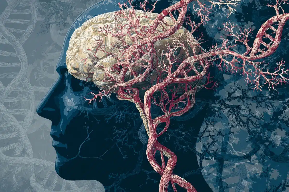
Long-term exposure to periodontal disease bacteria causes inflammation and degeneration of brain neurons in mice that is similar to the effects of Alzheimer’s disease in humans, according to a new study from researchers at the University of Illinois at Chicago.
The findings, which are published in PLOS ONE, suggest that periodontal disease, a common but preventable gum infection, may be an initiator of Alzheimer’s, which currently has no treatment or cure.
“Other studies have demonstrated a close association between periodontitis and cognitive impairment, but this is the first study to show that exposure to the periodontal bacteria results in the formation of senile plaques that accelerate the development of neuropathology found in Alzheimer’s patients,” said Dr. Keiko Watanabe, professor of periodontics at the UIC College of Dentistry and corresponding author on the study.
“This was a big surprise,” Watanabe said. “We did not expect that the periodontal pathogen would have this much influence on the brain, or that the effects would so thoroughly resemble Alzheimer’s disease.”
To study the impact of the bacteria on brain health, the Watanabe and her colleagues — including Dr. Vladimir Ilievski, UIC research assistant professor and co-author on the paper — established chronic periodontitis, which is characterized by soft tissue damage and bone loss in the oral cavity, in 10 wild-type mice. Another 10 mice served as the control group. After 22 weeks of repeated oral application of the bacteria to the study group, the researchers studied the brain tissue of the mice and compared brain health.
The researchers found that the mice chronically exposed to the bacteria had significantly higher amounts of accumulated amyloid beta — a senile plaque found in the brain tissue of Alzheimer’s patients. The study group also had more brain inflammation and fewer intact neurons due to degeneration.
These findings were further supported by amyloid beta protein analysis, and RNA analysis that showed greater expression of genes associated with inflammation and degeneration in the study group. DNA from the periodontal bacteria was also found in the brain tissue of mice in the study group, and a bacterial protein was observed inside their neurons.
“Our data not only demonstrate the movement of bacteria from the mouth to the brain, but also that chronic infection leads to neural effects similar to Alzheimer’s,” Watanabe said.
The researchers say these findings are powerful in part because they used a wild-type mouse model; most model systems used to study Alzheimer’s rely on transgenic mice, which have been genetically altered to more strongly express genes associated with the senile plaque and enable Alzheimer’s development.
“Using a wild-type mouse model added strength to our study because these mice were not primed to develop the disease, and use of this model gives additional weight to our findings that periodontal bacteria may kick-start the development of the Alzheimer’s,” Watanabe said.
The researchers say that understanding causality and risk factors for the development of Alzheimer’s is critical to the development of treatments, particularly when it comes to sporadic, or late-onset disease, which constitutes more than 95 percent of cases and has largely unknown causes and mechanisms.
While the findings are significant for the scientific community, Watanabe said there are lessons for everyone.
“Oral hygiene is an important predictor of disease, including diseases that happen outside the mouth,” she said. “People can do so much for their personal health by taking oral health seriously.”
Additional co-authors on the paper are Paulina Zuchowska, Stefan Green, Peter Toth, Michael Ragozzino, Khuong Le and Haider Aljewari of UIC, and Neil O’Brein-Simpson and Eric Reynolds of the University of Melbourne.
Source: Jackie Carey – University of Illinois
Publisher: Organized by NeuroscienceNews.com.
Image Source: NeuroscienceNews.com image is in the public domain.
Original Research: Open access research for “Chronic oral application of a periodontal pathogen results in brain inflammation, neurodegeneration and amyloid beta production in wild type mice” by Vladimir Ilievski, Paulina K. Zuchowska, Stefan J. Green, Peter T. Toth, Michael E. Ragozzino, Khuong Le, Haider W. Aljewari, Neil M. O’Brien-Simpson, Eric C. Reynolds, and Keiko Watanabe in PLOS ONE. Published October 3 2018.
doi:10.1371/journal.pone.0204941
[cbtabs][cbtab title=”MLA”]University of Illinois”Periodontal Disease Bacteria May Kick Start Alzheimer’s.” NeuroscienceNews. NeuroscienceNews, 4 October 2018.
<https://neurosciencenews.com/periodontal-disease-alzheimers-9955/>.[/cbtab][cbtab title=”APA”]University of Illinois(2018, October 4). Periodontal Disease Bacteria May Kick Start Alzheimer’s. NeuroscienceNews. Retrieved October 4, 2018 from https://neurosciencenews.com/periodontal-disease-alzheimers-9955/[/cbtab][cbtab title=”Chicago”]University of Illinois”Periodontal Disease Bacteria May Kick Start Alzheimer’s.” https://neurosciencenews.com/periodontal-disease-alzheimers-9955/ (accessed October 4, 2018).[/cbtab][/cbtabs]
Abstract
Chronic oral application of a periodontal pathogen results in brain inflammation, neurodegeneration and amyloid beta production in wild type mice
Background
The results from cross sectional and longitudinal studies show that periodontitis is closely associated with cognitive impairment (CI) and Alzhemer’s Disease (AD). Further, studies using animal model of periodontitis and human post-mortem brain tissues from subjects with AD strongly suggest that a gram-negative periodontal pathogen, Porphyromonas gingivalis (Pg) and/or its product gingipain is/are translocated to the brain. However, neuropathology resulting from Pg oral application is not known. In this work, we tested the hypothesis that repeated exposure of wild type C57BL/6 mice to orally administered Pg results in neuroinflammation, neurodegeneration, microgliosis, astrogliosis and formation of intra- and extracellular amyloid plaque and neurofibrillary tangles (NFTs) which are pathognomonic signs of AD.
Methods
Experimental chronic periodontitis was induced in ten wild type 8-week old C57BL/6 WT mice by repeated oral application (MWF/week) of Pg/gingipain for 22 weeks (experimental group). Another 10 wild type 8-week old C57BL/6 mice received vehicle alone (control group) MWF per week for 22 weeks. Brain tissues were collected and the presence of Pg/gingipain was determined by immunofluorescence (IF) microscopy, confocal microscopy, and quantitative PCR (qPCR). The hippocampi were examined for the signs of neuropathology related to AD: TNFα, IL1β, and IL6 expression (neuroinflammation), NeuN and Fluoro Jade C staining (neurodegeneration) and amyloid beta1-42 (Aβ42) production and phosphorylation of tau protein at Ser396 were assessed by IF and confocal microscopy. Further, gene expression of amyloid precursor protein (APP), beta-site APP cleaving enzyme 1 (BACE1), a disintegrin and metalloproteinase domain-containing protein10 (ADAM10) for α-secretase and presenilin1 (PSEN1) for ɣ-secretase, and NeuN (rbFox3) were determined by RT-qPCR. Microgliosis and astrogliosis were also determined by IF microscopy.
Results
Pg/gingipain was detected in the hippocampi of mice in the experimental group by immunohistochemistry, confocal microscopy, and qPCR confirming the translocation of orally applied Pg to the brain. Pg/gingipain was localized intra-nuclearly and peri-nuclearly in microglia (Iba1+), astrocytes (GFAP+), neurons (NeuN+) and was evident extracellularly. Significantly greater levels of expression of IL6, TNFα and IL1β were evident in experimental as compared to control group (p<0.01, p<0.00001, p<0.00001 respectively). In addition, microgliosis and astrogliosis were evident in the experimental but not in control group (p <0.01, p<0.0001 respectively). Neurodegeneration was evident in the experimental group based on a fewer number of intact neuronal cells assessed by NeuN positivity and rbFOX3 gene expression, and there was a greater number of degenerating neurons in the hippocampi of experimental mice assessed by Fluoro Jade C positivity. APP and BACE1 gene expression were increased in experimental group compared with control group (p<0.05, p<0.001 respectively). PSEN1 gene expression was higher in experimental than control group but the difference was not statistically significant (p = 0.07). ADAM10 gene expression was significantly decreased in experimental group compared with control group (p<0.01). Extracellular Aβ42 was detected in the parenchyma in the experimental but not in the control group (p< 0.00001). Finally, phospho-Tau (Ser396) protein was detected and NFTs were evident in experimental but not in the control group (p<0.00001).
Conclusions
This study is the first to show neurodegeneration and the formation of extracellular Aβ42 in young adult WT mice after repeated oral application of Pg. The neuropathological features observed in this study strongly suggest that low grade chronic periodontal pathogen infection can result in the development of neuropathology that is consistent with that of AD.