

Findings point to possible ways to fight some age-related diseases.
As we age, our body rhythms lose time before they finally stop. Breaking the body clock by genetically disrupting a core clock gene, Bmal1, in mice has long been known to accelerate aging , causing arthritis, hair loss, cataracts, and premature death.
New research now reveals that the nerve cells of these mice with broken clocks show signs of deterioration before the externally visible signs of aging are apparent, raising the possibility of novel approaches to staving off or delaying neurodegeneration – hallmarks of Parkinson’s and Alzheimer’s diseases.
Erik Musiek, M.D., Ph.D., who was a postdoctoral fellow in the lab of Garret FitzGerald, M.D., director of the Institute of Translational Medicine and Therapeutics, Perelman School of Medicine, University of Pennsylvania, took on this project four years ago. Musiek, now an assistant professor at Washington University, completed this line of research over the last two years in the lab of David Holtzman, M.D., also at WashU.
The Penn-WashU team found that the expression of certain clock genes, including Bmal1, plays a fundamental role in delaying emergence of age-related signs of decay in the brain. The clock proteins appear to do this by protecting the brain against oxidative stress – a process akin to rusting – that is normally controlled by enzymes that degrade harmful forms of oxygen generated in the course of normal metabolism. Their findings appear this week in the Journal of Clinical Investigation.
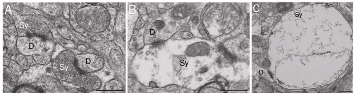
They found, to their surprise, that inflammation – reflected by activation of astrocytes – brain cells involved in this type of response, among other functions — was marked in young mice in which the clock was broken by deleting Bmal1. This anticipated even more marked changes in brain pathology as the mice aged, including declines in how parts of the brain connected to each other and degenerative features in nerve-cell anatomy – all characteristic of Parkinsons and Alzheimer’s disease in humans.
“When we saw this, we knew we were on to something,” notes Musiek.
Further experiments revealed that these effects were not restricted to disrupting the function of Bmal1, but also occurred when genes – Clock and Npas2 – with which Bmal1 works in tandem, were both removed. By contrast, deletion of other genes in the clock apparatus had no such effect.
As for mechanism, the exaggerated rusting, or oxidation, was key. Expression of several antioxidant enzymes, which normally keep oxidant stress in check are themselves controlled by clock proteins, and thus were depleted when the clock was broken. Musiek and his colleagues found evidence that inflammation and the attendant oxidant stress were both increased in the brains of the mutant mice.
Experimental drugs are beginning to emerge that may retain waning rhythms driven by the molecular clock. “Erik’s studies raise the intriguing possibility of novel therapeutic approaches to delaying the progress of age-related diseases, perhaps not only those related to the brain, as suggested by the present studies, but also in other systems, such as cardiometabolic function,” says FitzGerald.
In a final twist, the Penn-WashU team pinned the neuroprotective role of the body clock to clock genes in neurons and astrocytes, rather than changes in whole-animal circadian rhythms. By selectively deleting Bmal1 in these cell types, they found that the inflammatory aspects of astrocytes, neurodegeneration, and hallmarks of oxidative stress and inflammation seen when Bmal1 was missing in all cells of the body was recapitulated.
“Our findings indicate that the protein complex of BMAL1 with CLOCK or NPAS2, in addition to, or perhaps intrinsic, to the complex’s internal body-clock function, regulates protection of the brain from inflammation and oxygen free-radical induced damage. This dynamic system connects impaired clock-gene function to neurodegeneration for the first time,” says Musiek.
Notes about this neurodegeneration research
This study was supported by the National Institute of Neurological Disorders and Stroke and the National Heart, Lung, and Blood Institute (K08NS079405, R25NS065745, HL097800, P01NS074969, P30NS057105, NS056125)
Contact: Karen Kreeger – Penn Medicine
Source: Penn Medicine press release
Image Source: The image is credited to Erik Musiek, M.D., Ph.D., Journal of Clinical Investigation and is adapted from the Penn Medicine press release.
Original Research: Abstract for “Circadian clock proteins regulate neuronal redox homeostasis and neurodegeneration” by Erik S. Musiek, Miranda M. Lim, Guangrui Yang, Adam Q. Bauer, Laura Qi, Yool Lee, Jee Hoon Roh, Xilma Ortiz-Gonzalez, Joshua T. Dearborn, Joseph P. Culver, Erik D. Herzog, John B. Hogenesch, David F. Wozniak, Krikor Dikranian, Benoit I. Giasson, David R. Weaver, David M. Holtzman and Garret A. FitzGerald in Journal of Clinical Investigation. Published online November 15 2013 doi:10.1172/JCI70317


