

Specific cardiovascular risk factors, such as alcohol consumption, smoking, obesity and diabetes, are associated with smaller regional brain volumes that may be early indicators of Alzheimer’s disease and dementia according to a study published online in the journal Radiology.
“We already know that vascular risk factors damage the brain and can result in cognitive impairment,” said Kevin S. King, M.D., assistant professor of radiology at the Keck School of Medicine of the University of Southern California in Los Angeles. “But our findings give us a more concrete idea about the relationship between specific vascular risk factors and brain health.”
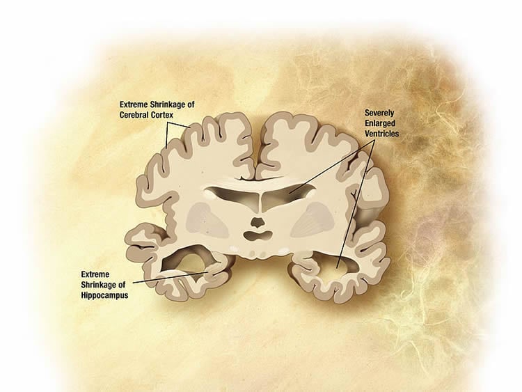
Prior studies have linked cardiovascular risk factors and cognitive decline, but the new study focused on specific risk factors and examined three main brain regions, including the hippocampus, precuneus and posterior cingulate cortex. Because of each region’s connection to memory retrieval, gray matter volume loss in these areas may be a predictor of Alzheimer’s disease and dementia.
In the new study, Dr. King and colleagues analyzed results from 1,629 individuals in the Dallas Heart Study (DHS) and divided the participants into two age groups. There were 805 participants under age 50, and 824 age 50 and older. The researchers evaluated the participants’ data from the initial baseline visit, which included laboratory and clinical analysis, and the follow-up visit seven years later consisting of a brain MRI and cognitive test, measuring mild cognitive impairment and preclinical Alzheimer’s disease.
By comparing the initial visit in which cardiovascular risk factors were identified to the MRI results and cognitive scores, the team was able to distinguish the specific risk factors of alcohol consumption, smoking, diabetes, and obesity and their relationship to smaller volumes in the three targeted regions of the brain. The results confirmed that lower cognitive test scores correlated with lower brain volumes in each area.
The study found that risk factors of alcohol use and diabetes were associated with smaller total brain volume, while smoking and obesity were linked with reduced volumes of the posterior cingulate cortex, the area of the brain connected with memory retrieval as well as emotional and social behavior. In addition, lower hippocampal mass was linked to both alcohol consumption and smoking whereas alcohol use, obesity and high fasting blood glucose numbers correlated with reduced precuneus size.
The findings also suggest that in patients age 50 and older, diminished hippocampal and precuneus volumes may be early risk indicators for cognitive decline, while smaller posterior cingulate volumes are better predictors in patients under age 50.
Dr. King believes that additional studies can provide the ability to better identify the impact of specific cardiovascular risk factors on the brain and improve patient understanding of brain diseases.
“We currently do not have effective treatments for Alzheimer’s disease, so the focus is on prevention,” he said. “In the future, we may be able to provide patients with useful and actionable information about the impact different risk factors may be having on their brain health during routine clinical imaging. And since no special imaging equipment is needed, there is a great potential to provide this service at many centers across the country.”
Funding: This research was supported by the NIH.
Source: Linda Brooks – RSNA
Image Credit: The image is in the public domain
Original Research: Abstract for “Cardiovascular Risk Factors Associated with Smaller Brain Volumes in Regions Identified as Early Predictors of Cognitive Decline” by Rajiv N. Srinivasa, Heidi C. Rossetti, Mohit K. Gupta, Roger N. Rosenberg, Myron F. Weiner, Ronald M. Peshock, Roderick W. McColl, Linda S. Hynan, Richard T. Lucarelli, and Kevin S. King in Radiology. Published online July 28 2015 doi:10.1148/radiol.2015142488
Abstract
Cardiovascular Risk Factors Associated with Smaller Brain Volumes in Regions Identified as Early Predictors of Cognitive Decline
Purpose
To determine in a large multiethnic cohort the cardiovascular and genetic risk factors associated with smaller volume in the hippocampus, precuneus, and posterior cingulate, and their association with preclinical deficits in cognitive performance in patients younger and older than 50 years.
Materials and Methods
The institutional review board approved the study and all participants provided written informed consent. Eligible for this study were 1629 participants (700 men and 929 women; mean age, 50.0 years ± 10.2 [standard deviation]) drawn from the population-based Dallas Heart Study who underwent laboratory and clinical analysis in an initial baseline visit and approximately 7 years later underwent brain magnetic resonance imaging with automated volumetry and cognitive assessment with the Montreal Cognitive Assessment (MoCA). Regression analysis showed associations between risk factors and segmental volumes, and associations between these volumes with cognitive performance in participants younger and older than 50 years.
Results
Lower hippocampal volume was associated with previous alcohol consumption (standardized estimate, −0.04; P = .039) and smoking (standardized estimate, −0.04; P = .048). Several risk factors correlated with lower total brain, posterior cingulate, and precuneus volumes. Higher total (standardized estimate, 0.06; P = .050), high-density lipoprotein (standardized estimate, 0.07; P = .003), and low-density lipoprotein (standardized estimate, 0.04; P = .037) cholesterol levels were associated with larger posterior cingulate volume, and higher triglyceride levels (standardized estimate, 0.06; P = .004) were associated with larger precuneus volume. Total MoCA score was associated with posterior cingulate volume (standardized estimate, 0.13; P = .001) in younger individuals and with hippocampal (standardized estimate, 0.06; P < .05) and precuneus (standardized estimate, 0.08; P < .023) volumes in older adults. Conclusion
Smaller volumes in specific brain regions considered to be early markers of dementia risk were associated with specific cardiovascular disease risk factors and cognitive deficits in a predominantly midlife multiethnic population-based sample. Additionally, the risk factors most associated with these brain volumes differed in participants younger and older than 50 years, as did the association between brain volume and MoCA score.
“Cardiovascular Risk Factors Associated with Smaller Brain Volumes in Regions Identified as Early Predictors of Cognitive Decline” by Rajiv N. Srinivasa, Heidi C. Rossetti, Mohit K. Gupta, Roger N. Rosenberg, Myron F. Weiner, Ronald M. Peshock, Roderick W. McColl, Linda S. Hynan, Richard T. Lucarelli, and Kevin S. King in Radiology. Published online July 28 2015 doi:10.1148/radiol.2015142488


