

Summary: Participating in exercise improved synaptic pruning in mouse models of autism. The study also found microglia dependant synaptic pruning is impaired by maternal inflammation, which has been previously connected to the development of ASD.
Source: University of Tokyo
A mouse model of autism has revealed how exercise changes the structure of the brain by eliminating the excess connections between neurons characteristic of autism spectrum disorder (ASD). After one month of voluntary running on a wheel, behavioral differences disappeared and structural differences in ASD model mice’s brains were reduced.
“Voluntary exercise can be a safe, drug-free way to improve overall health and now we have a better understanding of how exercise can positively change brain structure in ASD,” said Associate Professor Ryuta Koyama, who led the research team at the University of Tokyo.
This research study was performed in mice and researchers caution against expecting exercise to act as a panacea for humans.
“A very important part of this research is that the mice were not forced to exercise – any physical activity was voluntary. If children are interested, they should be encouraged to do any exercise they want, but this research does not reveal how different activities might affect the brains of children with ASD,” said Koyama.
Fitness for the mind
About a decade ago, small trials in children began showing that regular exercise could improve the social difficulties and repetitive behaviors common in ASD. Although many studies investigated how exercise can make large-scale changes to already healthy brains or postpone age-related neurodegeneration, there were no studies on the fine-tuned changes that exercise might make in brains with ASD.
Although the brains of healthy children naturally prune away redundant connections between neurons as they age, a defining feature of the brains of people with ASD is too many connections.
The results from the mouse model study indicate that exercise may somehow activate the brain’s normal pruning processes.
“Exercise likely enhanced the contrast between active and inactive neuronal connections so that the weak ones could be more easily targeted for removal,” said Koyama.
A gym for mice
ASD model mice display lifelong behaviors of neurodevelopmental difference: They interact less with new mice, they groom themselves repetitively, and they take longer to start eating when in a new environment, a sign of anxiety.
Researchers placed a running wheel in the corner of the ASD model mice’s cages so they could run as much as they wished. Mice had access to the wheel from the time they were four weeks old until eight weeks old. Mice are adults at eight weeks old.
“Part of the variability of our data is that we could not give all mice the same amount of exercise. Some mice may have exercised just a little, some may have exercised very intensely,” said Koyama.
Despite wide variation between individual mice, overall, ASD model mice that could exercise showed behavior that was similar to healthy mice after just 30 days of access to the running wheel.
Cellular studies for a closer look
Researchers performed additional experiments to look inside the brains of ASD model mice.
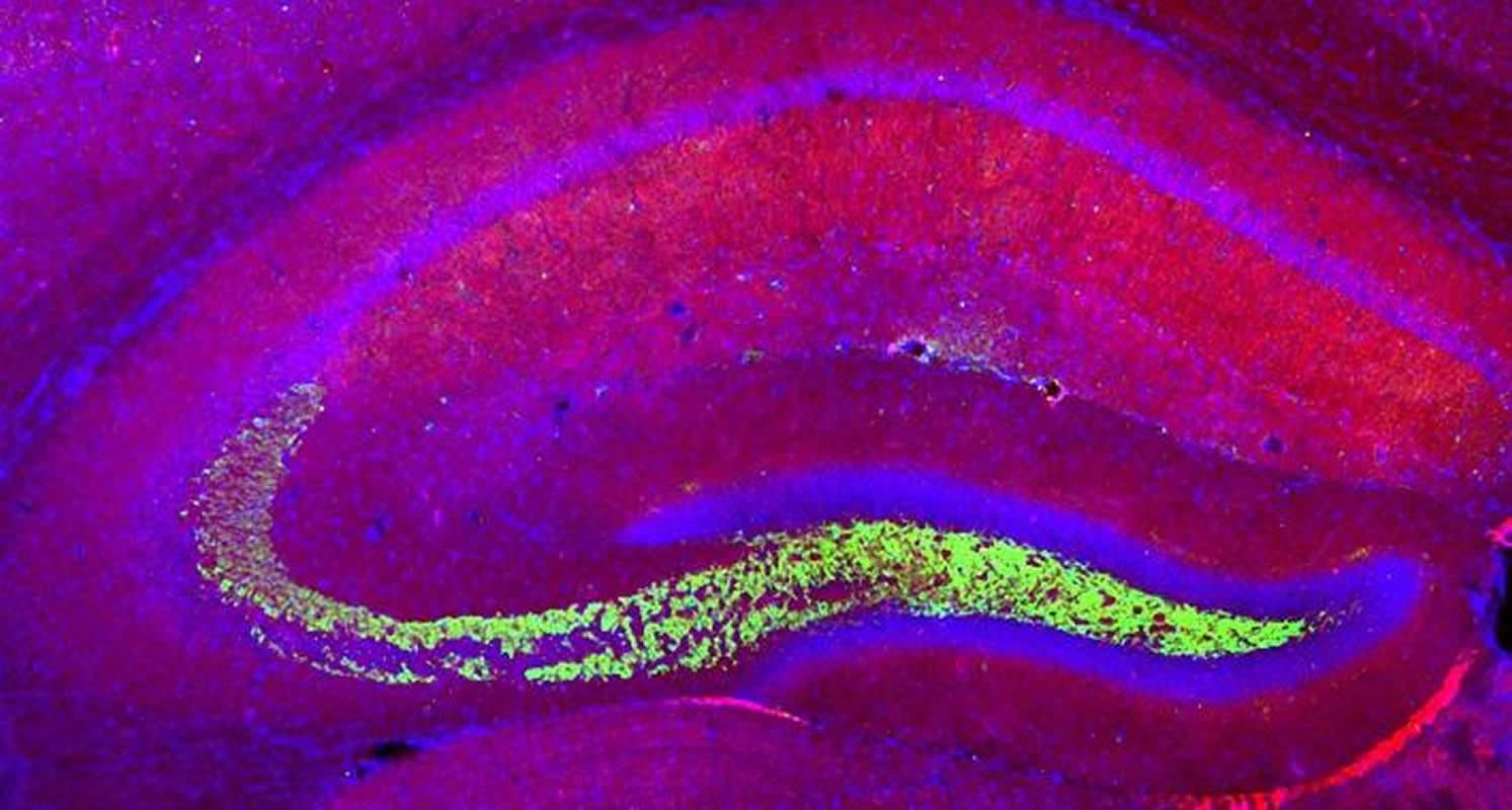
Specifically, researchers focused on a structure called mossy fibers in the hippocampus, an area of the brain thought to be important for memory and a source of new neurons (neurogenesis) in adults. Individual neurons can be connected to dozens of mossy fibers sent out by other neurons.
Immune cells called microglia change brain structure by engulfing and digesting less active synapses, which is why they are sometimes referred to as “the brain’s garbage collectors.”
Researchers used molecular genetics and fluorescent imaging techniques to identify which mossy fibers were most active and saw that microglia spared those active synapses, while removing the less active ones nearby.
Researchers also connected microglia to synaptic pruning in healthy mice. Juvenile mice without ASD were given medication to prevent normal microglia activity. After just five days of medication, researchers detected significant differences in the density of brain synapses between mice whose microglia were inhibited and their unmedicated peers.
Although the causes of ASD are an area of active research, Koyama’s research team states that the reversible aspects of ASD and the connection between exercise and microglia should also receive continued attention.
Source:
University of Tokyo
Media Contacts:
Ryuta Koyama – University of Tokyo
Image Source:
The image is credited to Ryuta Koyama.
Original Research: Open access
“Exercise reverses behavioral and synaptic abnormalities after maternal inflammation”. Andoh M, Shibata K, Okamoto K, Onodera J, Morishita K, Miura Y, Ikegaya Y, Koyama R.
Cell Reports. doi:10.1016/j.celrep.2019.05.015
Abstract
Exercise reverses behavioral and synaptic abnormalities after maternal inflammation
Highlights
• MIA causes behavioral and synaptic abnormalities in the offspring
• Microglia-dependent synaptic engulfment is impaired by MIA
• Voluntary running in adulthood ameliorates MIA-induced abnormalities
• Voluntary running stimulates microglia-mediated engulfment of surplus synapses
Summary
Abnormal behaviors in individuals with neurodevelopmental disorders are generally believed to be irreversible. Here, we show that voluntary wheel running ameliorates the abnormalities in sociability, repetitiveness, and anxiety observed in a mouse model of a neurodevelopmental disorder induced by maternal immune activation (MIA). Exercise activates a portion of dentate granule cells, normalizing the density of hippocampal CA3 synapses, which is excessive in the MIA-affected offspring. The synaptic surplus in the MIA offspring is induced by deficits in synapse engulfment by microglia, which is normalized by exercise through microglial activation. Finally, chemogenetically induced activation of granule cells promotes the engulfment of CA3 synapses. Thus, our study proposes a role of voluntary exercise in the modulation of behavioral and synaptic abnormalities in neurodevelopmental disorders.


