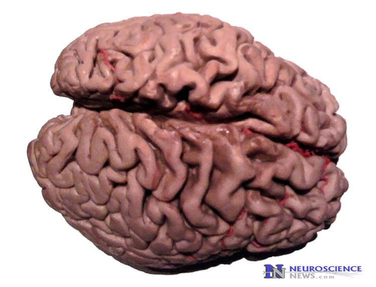
Summary: Researchers discover a decline in glucose levels in the brain that occurs during the early stages of MCI.
Source: Temple University Health System.
One of the earliest signs of Alzheimer’s disease is a decline in glucose levels in the brain. It appears in the early stages of mild cognitive impairment — before symptoms of memory problems begin to surface. Whether it is a cause or consequence of neurological dysfunction has been unclear, but new research at the Lewis Katz School of Medicine at Temple University now shows unequivocally that glucose deprivation in the brain triggers the onset of cognitive decline, memory impairment in particular.
“In recent years, advances in imaging techniques, especially positron emission tomography (PET), have allowed researchers to look for subtle changes in the brains of patients with different degrees of cognitive impairment,” explained Domenico Praticò, MD, Professor in the Center for Translational Medicine at the Lewis Katz School of Medicine at Temple University (LKSOM). “One of the changes that has been consistently reported is a decrease in glucose availability in the hippocampus.”
The hippocampus plays a key role in processing and storing memories. It and other regions of the brain, however, rely exclusively on glucose for fuel — without glucose, neurons starve and eventually die.
The new study, published online January 31 in the journal Translational Psychiatry, is the first to directly link memory impairment to glucose deprivation in the brain specifically through a mechanism involving the accumulation of a protein known as phosphorylated tau.
“Phosphorylated tau precipitates and aggregates in the brain, forming tangles and inducing neuronal death,” Dr. Praticò explained. In general, a greater abundance of neurofibrillary tau tangles is associated with more severe dementia.
The study also is the first to identify a protein known as p38 as a potential alternate drug target in the treatment of Alzheimer’s disease. Neurons activate p38 protein in response to glucose deprivation, possibly as a defensive mechanism. In the long run, however, its activation increases tau phosphorylation, making the problem worse.
To investigate the impact of glucose deprivation on the brain, Dr. Praticò’s team used a mouse model that recapitulates memory impairments and tau pathology in Alzheimer’s disease. At about 4 or 5 months of age, some of the animals were treated with 2-deoxyglucose (DG), a compound that stops glucose from entering and being utilized by cells. The compound was administered to the mice in a chronic manner, over a period of several months. The animals were then evaluated for cognitive function. In a series of maze tests to assess memory, glucose-deprived mice performed significantly worse than their untreated counterparts.
When examined microscopically, neurons in the brains of DG-treated mice exhibited abnormal synaptic function, suggesting that neural communication pathways had broken down. Of particular consequence was a significant reduction in long-term potentiation- – the mechanism that strengthens synaptic connections to ensure memory formation and storage.
Upon further examination, the researchers discovered high levels of phosphorylated tau and dramatically increased amounts of cell death in the brains of glucose-deprived mice. To find out why, Dr. Praticò turned to p38, which in earlier work his team had identified as a driver of tau phosphorylation. In the new study, they found that memory impairment was directly associated with increased p38 activation.
“The findings are very exciting,” Dr. Praticò said. “There is now a lot of evidence to suggest that p38 is involved in the development of Alzheimer’s disease.”

The findings also lend support to the idea that chronically occurring, small episodes of glucose deprivation are damaging for the brain. “There is a high likelihood that those types of episodes are related to diabetes, which is a condition in which glucose cannot enter the cell,” he explained. “Insulin resistance in type 2 diabetes is a known risk factor for dementia.”
According to Dr. Praticò, the next step is to inhibit p38 to see if memory impairments can be alleviated, despite glucose deprivation. “It is an exciting avenue of research. A drug targeting this protein could bring big benefits for patients,” he added.
Other researchers involved in the new study include Elisabetta Lauretti, Jian-Guo Li, and Antonio Di Meco in the Department of Pharmacology and Center for Translational Medicine at LKSOM.
Funding: The research was supported in part by a grant from the Wanda Simone Endowment Fund for Neuroscience.
Source: Jeremy Walter – Temple University Health System
Image Source: NeuroscienceNews.com image is in the public domain
Original Research: Full open access research for “Glucose deficit triggers tau pathology and synaptic dysfunction in a tauopathy mouse model” by E Lauretti, J-G Li, A Di Meco & D Praticò in Translational Psychiatry. Published online January 31 2017 doi:10.1038/tp.2016.296
[cbtabs][cbtab title=”MLA”]Temple University Health System “Glucose Deprivation in the Brain Sets Stage for Alzheimer’s.” NeuroscienceNews. NeuroscienceNews, 31 January 2017.
<https://neurosciencenews.com/alzheimers-glucose-deprivation-6035/>.[/cbtab][cbtab title=”APA”]Temple University Health System (2017, January 31). Glucose Deprivation in the Brain Sets Stage for Alzheimer’s. NeuroscienceNew. Retrieved January 31, 2017 from https://neurosciencenews.com/alzheimers-glucose-deprivation-6035/[/cbtab][cbtab title=”Chicago”]Temple University Health System “Glucose Deprivation in the Brain Sets Stage for Alzheimer’s.” https://neurosciencenews.com/alzheimers-glucose-deprivation-6035/ (accessed January 31, 2017).[/cbtab][/cbtabs]
Abstract
Glucose deficit triggers tau pathology and synaptic dysfunction in a tauopathy mouse model
Clinical investigations have highlighted a biological link between reduced brain glucose metabolism and Alzheimer’s disease (AD). Previous studies showed that glucose deprivation may influence amyloid beta formation in vivo but no data are available on the effect that this condition might have on tau protein metabolism. In the current paper, we investigated the effect of glucose deficit on tau phosphorylation, memory and learning, and synaptic function in a transgenic mouse model of tauopathy, the h-tau mice. Compared with controls, h-tau mice with brain glucose deficit showed significant memory impairments, reduction of synaptic long-term potentiation, increased tau phosphorylation, which was mediated by the activation of P38 MAPK Kinase pathway. We believe our studies demonstrate for the first time that reduced glucose availability in the central nervous system directly triggers behavioral deficits by promoting the development of tau neuropathology and synaptic dysfunction. Since restoring brain glucose levels and metabolism could afford the opportunity to positively influence the entire AD phenotype, this approach should be considered as a novel and viable therapy for preventing and/or halting the disease progression.
“Glucose deficit triggers tau pathology and synaptic dysfunction in a tauopathy mouse model” by E Lauretti, J-G Li, A Di Meco & D Praticò in Translational Psychiatry. Published online January 31 2017 doi:10.1038/tp.2016.296


