

Summary: Episodic exposure to nicotine, caffeine, and amphetamines trigger malfunctions in the fetal brain, specifically affecting the development of the indusium griseum.
Source: Medical University of Vienna
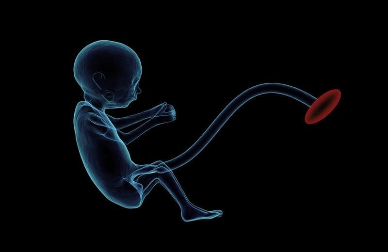
If pregnant women take significant amounts of the psychostimulants coffee, nicotine, and amphetamine during pregnancy, their children have a higher risk of developing neurological and psychiatric problems later in life. Researchers at MedUni Vienna’s Center for Brain Research have now successfully identified the regions of the brain that act as “hot spots” for psychostimulants and discovered that the mother’s reactions to these substances are substantially different from those of their baby. This study has now been published in the multidisciplinary journal PNAS.
Drug abuse during pregnancy carries considerable risk and negatively impacts fetal development. Even though the mother does not react particularly strongly to certain psychostimulants, these drugs can nonetheless permanently affect brain development of her baby or child.
The precise areas of the brain that are affected by maternal drug consumption were hitherto unknown. The recent study conducted by MedUni Vienna’s Center for Brain Research, working in conjunction with the Swedish Karolinska Institute, has now shown that episodic exposure to amphetamine, nicotine or caffeine during pregnancy triggers an extensive malfunction in the fetal brain, which specifically affects the development of the indusium griseum (IG). The IG is a cerebral area that reacted to all psychostimulants tested in a mouse model.
“In the indusium griseum, we found a new type of neuron that is affected by psychostimulants, in that they greatly inhibit its development so that the baby is born with neurons still in a fetal-like state. A major consequence of this is that these cells are no longer able to integrate appropriately into the brain in the long term,” explains principal investigator Tibor Harkany from MedUni Vienna’s Center for Brain Research.
For their analysis, the researchers combined conventional neuroanatomy with the very latest RNA sequencing techniques to show how molecular impairments occur in neurons of the indusium griseum. “In particular, the level of a certain protein, secretagogin, is reduced. This deficiency impairs the mechanism by which neurons are able to process information. This has also been proven in genetic models. Mice that do not have this protein respond to psychostimulants such as methamphetamine more strongly and with an increased risk of developing epilepsy,” explains lead author of the study Janos Fuzik from MedUni Vienna’s Center for Brain Research. The result: Children could also develop an increased risk of neurological complications later on, because the anatomical structure of the indusium griseum is also present as a thin layer of gray matter in the human brain.
Neuronal population in the human IG documented for the first time
According to Harkany, a “surprising observation” was that there is any neuronal population in the indusium griseum of men at all. “Up until now, science believed that there are no neurons, or only a tiny numbers of neurons, in this area,” he says. Whether the function of these neurons in the human brain is equivalent to those of mice needs further study, though.
“However, this at least shows that brain networks are more complex than previously thought, and that coordination of brain functions is much more diverse than we might have expected,” says Thomas Hokfelt of the Karolinska Institute, who is an adjunct professor at MedUni Vienna’s Center for Brain Research. Since these neurons are involved in cognitive networks and probably facilitate cognition, when their networks become damaged by psychostimulants during their developmental phase, life-long deficiencies can be expected.
Source:
Medical University of Vienna
Media Contacts:
Tibor Harkany – Medical University of Vienna
Image Source:
The image is in the public domain.
Original Research: Closed access
“Brain-wide genetic mapping identifies the indusium griseum as a prenatal target of pharmacologically unrelated psychostimulants”. Janos Fuzik et al.
PNAS doi:10.1073/pnas.1904006116.
Abstract
Brain-wide genetic mapping identifies the indusium griseum as a prenatal target of pharmacologically unrelated psychostimulants
Psychostimulant use is an ever-increasing socioeconomic burden, including a dramatic rise during pregnancy. Nevertheless, brain-wide effects of psychostimulant exposure are incompletely understood. Here, we performed Fos-CreERT2–based activity mapping, correlated for pregnant mouse dams and their fetuses with amphetamine, nicotine, and caffeine applied acutely during midgestation. While light-sheet microscopy-assisted intact tissue imaging revealed drug- and age-specific neuronal activation, the indusium griseum (IG) appeared indiscriminately affected. By using GAD67gfp/+ mice we subdivided the IG into a dorsolateral domain populated by γ-aminobutyric acidergic interneurons and a ventromedial segment containing glutamatergic neurons, many showing drug-induced activation and sequentially expressing Pou3f3/Brn1 and secretagogin (Scgn) during differentiation. We then combined Patch-seq and circuit mapping to show that the ventromedial IG is a quasi-continuum of glutamatergic neurons (IG-Vglut1+) reminiscent of dentate granule cells in both rodents and humans, whose dendrites emanate perpendicularly toward while their axons course parallel with the superior longitudinal fissure. IG-Vglut1+ neurons receive VGLUT1+ and VGLUT2+ excitatory afferents that topologically segregate along their somatodendritic axis. In turn, their efferents terminate in the olfactory bulb, thus being integral to a multisynaptic circuit that could feed information antiparallel to the olfactory–cortical pathway. In IG-Vglut1+ neurons, prenatal psychostimulant exposure delayed the onset of Scgn expression. Genetic ablation of Scgn was then found to sensitize adult mice toward methamphetamine-induced epilepsy. Overall, our study identifies brain-wide targets of the most common psychostimulants, among which Scgn+/Vglut1+ neurons of the IG link limbic and olfactory circuits.


