Summary: Neurofilament light protein in plasma may be a noninvasive biomarker for neurodegeneration associated with Alzheimer’s disease.
Source: Lund University
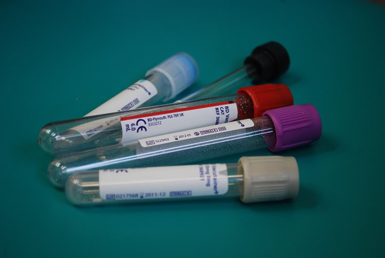
A new study confirms that a simple blood test can reveal whether there is accelerating nerve cell damage in the brain. The researchers analysed neurofilament light protein (NFL) in blood samples from patients with Alzheimer’s disease. Recently published in JAMA Neurology, the study suggests that the NFL concentration in the blood could be able to indicate if a drug actually affects the loss of nerve cells.
The blood samples were collected over several years, and on multiple occasions, from 1 182 patients with different degrees of cognitive impairment, and 401 healthy subjects in a control group.
Very sensitive methods have been developed in recent years to measure the presence of certain substances in the blood that can indicate damage in the brain and neurological diseases such as Parkinson’s, multiple sclerosis (MS) and Alzheimer’s. Neurofilament light protein (NFL) is one such substance.
“Standard methods for indicating nerve cell damage involve measuring the patient’s level of certain substances using a lumbar puncture or examining a brain MRI. These methods are complicated, take time and are costly. Measuring NFL in the blood can be cheaper and is also easier for the patient”, explains Niklas Mattsson, a researcher at Lund University and physician at Skåne University Hospital, who led the study.
When nerve cells in the brain are damaged or die, NFL protein leaks into the cerebrospinal fluid and onwards into the blood. It was previously known that the levels of NFL are elevated among people with neurodegenerative diseases, but there has been a lack of long-term studies.
“We discovered that the NFL concentration increases over time in Alzheimer’s disease and that these elevated levels also are in line with the accumulated brain damage, which we can measure using lumbar punctures or magnetic resonance imaging”, says Niklas Mattsson.
The new study focuses on the common form of the disease, sporadic Alzheimer’s disease. It is one of the most widespread chronic diseases in the world and the most common cause of dementia. The researchers have analysed a large number of blood samples collected over several years from a total of 1 583 patients.
“A recently published small-scale German-American study presented similar results on familial Alzheimer’s disease, a very rare form of the disease that is strongly related to heredity. Taken together, these studies indicate that NFL in the blood can be used to measure damage to brain cells in various forms of Alzheimer’s disease”, says Niklas Mattsson.
Alzheimer’s is a complex, difficult-to-diagnose disease that develops gradually. The disease involves the deterioration of cognitive and physical functions along with the atrophy and death of brain cells. At present, there is no treatment that can reduce the loss of nerve cells in the brain. Drugs are available to mitigate cognitive disorders, but not to slow the course of the disease.

Measurements of the NFL concentration in the blood could indicate if a medicine is actually affecting the loss of nerve cells, when an optimal dosage of the drug has been reached or if another drug should be tried.
“Within drug development, it can be valuable to detect the effects of the trialled drug at an early stage and to be able to test on people who do not yet have full-blown Alzheimer’s”, says Niklas Mattsson and continues;
“In previous drug trials there has been considerable uncertainty about the effects of the drugs. There are several reasons for this. For example, some of the patients involved probably did not have Alzheimer’s disease. In other cases, is was unclear if the drug had been introduced too late in the course of the disease. Measuring the NFL concentration in the blood could make things easier for future drug development, both through following the effects of the drug and by including test subjects who display markers of nerve cell deterioration. This approach will enable more reliable conclusions to be drawn from the results”.
Niklas Mattsson emphasises the importance of continuing to examine how sensitive the measurement of NFL in the blood is as a marker for Alzheimer’s disease and what can be expected from longitudinal changes. The effects of promising drugs also need to be confirmed in new drug studies.
However, he believes that the method is not far from becoming a standard clinical procedure.
“Preparatory work is ongoing at Sahlgrenska University Hospital in Gothenburg to make this method available as a clinical procedure in the near future. Physicians can then use the method to measure damage to nerve cells in Alzheimer’s disease and other brain disorders through a simple blood test”, Niklas Mattsson concludes.
Source:
Lund University
Media Contacts:
Niklas Mattsson – Lund University
Image Source:
The image is in the public domain.
Original Research: Closed access.
“Association Between Longitudinal Plasma Neurofilament Light and Neurodegeneration in Patients With Alzheimer Disease” Niklas Mattsson et al. JAMA Neurology. doi:10.1001/jamaneurol.2019.0765
Abstract
Association Between Longitudinal Plasma Neurofilament Light and Neurodegeneration in Patients With Alzheimer Disease
Importance Plasma neurofilament light (NfL) has been suggested as a noninvasive biomarker to monitor neurodegeneration in Alzheimer disease (AD), but studies are lacking.
Objective To examine whether longitudinal plasma NfL levels are associated with other hallmarks of AD.
Design, Setting, and Participants This North American cohort study used data from 1583 individuals in the multicenter Alzheimer’s Disease Neuroimaging Initiative study from September 7, 2005, through June 16, 2016. Patients were eligible for inclusion if they had NfL measurements. Annual plasma NfL samples were collected for up to 11 years and were analyzed in 2018.
Exposures Clinical diagnosis, Aβ and tau cerebrospinal fluid (CSF) biomarkers, imaging measures (magnetic resonance imaging and fluorodeoxyglucose–positron emission tomography), and tests on cognitive scores.
Main Outcomes and Measures The primary outcome was the association between baseline exposures (diagnosis, CSF biomarkers, imaging measures, and cognition) and longitudinal plasma NfL levels, analyzed by an ultrasensitive assay. The secondary outcomes were the associations between a multimodal classification scheme with Aβ, tau, and neurodegeneration (ie, the ATN system) and plasma NfL levels and between longitudinal changes in plasma NfL levels and changes in the other measures.
Results Of the included 1583 participants, 716 (45.2%) were women, and the mean (SD) age was 72.9 (7.1) years; 401 had cognitive impairment, 855 had mild cognitive impairment, and 327 had AD dementia. The NfL level was increased at baseline in patients with mild cognitive impairment and AD dementia (mean levels: cognitive unimpairment, 32.1 ng/L; mild cognitive impairment, 37.9 ng/L; and AD dementia, 45.9 ng/L; P < .001) and increased in all diagnostic groups, with the greatest increase in patients with AD dementia. A longitudinal increase in NfL level correlated with baseline CSF biomarkers (low Aβ42 [P = .001], high total tau [P = .02], and high phosphorylated tau levels [P = .02]), magnetic resonance imaging measures (small hippocampal volumes [P < .001], thin regional cortices [P = .009], and large ventricular volumes [P = .002]), low fluorodeoxyglucose–positron emission tomography uptake (P = .01), and poor cognitive performance (P < .001) for a global cognitive score. With use of the ATN system, increased baseline NfL levels were seen in A–T+N+ (P < .001), A+T–N+ (P < .001), and A+T+N+ (P < .001), and increased rates of NfL levels were seen in A–T+N– (P = .009), A–T+N+ (P = .02), A+T–N+ (P = .04), and A+T+N+ (P = .002). Faster increase in NfL levels correlated with faster increase in CSF biomarkers of neuronal injury, faster rates of atrophy and hypometabolism, and faster worsening in global cognition (all P < .05 in patients with mild cognitive impairment; associations differed slightly in cognitively unimpaired controls and patients with AD dementia).
Conclusions and Relevance The findings suggest that plasma NfL can be used as a noninvasive biomarker associated with neurodegeneration in patients with AD and may be useful to monitor effects in trials of disease-modifying drugs.


