

Summary: A research team demonstrated that the loss of healthy TDP-43 function specifically within microglia throws the entire brain’s developmental pipeline into chaos. Without functional TDP-43, microglial cells suffer a severe breakdown of the TREM2–DAP12 signaling pathway, stripping them of their ability to clear away routine structural myelin errors. This neuro-immunological breakdown triggers a cascading failure that disrupts oligodendrocyte health and culminates in severe adult motor deficits.
Key Facts
- The Guardians of Development: Microglia are tasked with acting as the brain’s frontline defense and structural maintenance crew. During early brain development, they routinely clear out dead cells, fight local infections, and prune cellular debris to ensure structural efficiency.
- The Glial Cross-Talk Failure: When the team deleted TDP-43 inside early-stage microglia, the downstream damage rippled across entirely different cell lines. Advanced imaging revealed structural flaws in early brain development, coupled with severe abnormalities in myelin, the protective insulating sheath wrapped around nerve fibers.
- Oligodendrocyte Collateral Damage: This structural disruption directly impacted oligodendrocytes, the highly specialized glial cells responsible for manufacturing and repairing myelin sheaths. The loss of TDP-43 in microglia altered the molecular environment so severely that oligodendrocytes fell into a state of cellular dysfunction.
- The TREM2–DAP12 Disruption: Under normal conditions, the brain naturally generates minor myelin errors during the intense wiring phases of early life. Microglia rely on a specialized signaling pathway known as the TREM2–DAP12 axis to detect, engulf, and clean up these routine debris anomalies. Without functional TDP-43, this vital pathway breaks down completely.
- The Adult Motor Consequence: Left unchecked, the accumulation of raw myelin debris permanently harms neural transmission pathways. Mice that lacked microglial TDP-43 during childhood grew up to display striking, highly measurable physical motor impairments and behavioral deficits as adults.
Source: University of Lausanne
Rosa Chiara Paolicelli’s team at the Department of Biomedical Sciences of Unil uncovered a new role for the TDP-43 protein in controlling the function of microglia, the immune cells of the brain. Their study shows how the loss of this protein in microglia may contribute to the development of neurological diseases.
The biology of microglia lies at the heart of the research conducted by Rosa Chiara Paolicelli’s group, Associate Professor in the Department of Biomedical Sciences at the Faculty of Biology and Medicine of the University of Lausanne (Unil). While microglia are considered the primary defense of the central nervous system, they are also involved in various neurodegenerative diseases. In their latest study, the Lausanne-based team demonstrated how dysfunctional microglia can disrupt brain function and lead to motor deficits.
The study was published in the 8 July 2026 issue of Nature Neuroscience.
The Brain’s “Guardians”
Microglia are small cells belonging to the glial cell family that reside in the central nervous system (the brain, spinal cord, and retina). They are the principal immune cells of the brain, often referred to as its “guardians.” In addition to destroying pathogenic foreign agents and fighting infections, they are also responsible for clearing dead cells and other cellular debris.
The Unil neuroscientists, in collaboration with colleagues from several Swiss and international institutions, focused their attention on the protein TDP-43. This protein is known to play a role in several neurodegenerative diseases, including amyotrophic lateral sclerosis (ALS) and certain forms of dementia. In these disorders, TDP-43, which is normally located in the cell nucleus, becomes mislocalized to the cytoplasm, where it forms abnormal aggregates.
Credit: Neuroscience News
The progressive loss of the protein from the nucleus, where it normally performs its essential functions, combined with the toxicity of the cytoplasmic aggregates, results in a loss of function and disrupts normal cellular activity. While this phenomenon has been extensively studied in neurons, its impact on microglia remains poorly understood.
“In our study, we sought to better understand what happens when TDP-43 is no longer functional in microglia, using the mouse as an experimental model,” explains Rosa Chiara Paolicelli, who led the research. “To achieve this, we genetically deleted the gene encoding this protein specifically in microglial cells. We observed that mice lacking TDP-43 in their microglia from early life stages developed motor impairments in adulthood.”
Harmful Effects at Multiple Levels
Using a range of imaging techniques, the researchers revealed that TDP-43 dysfunction affects the brain at several levels.
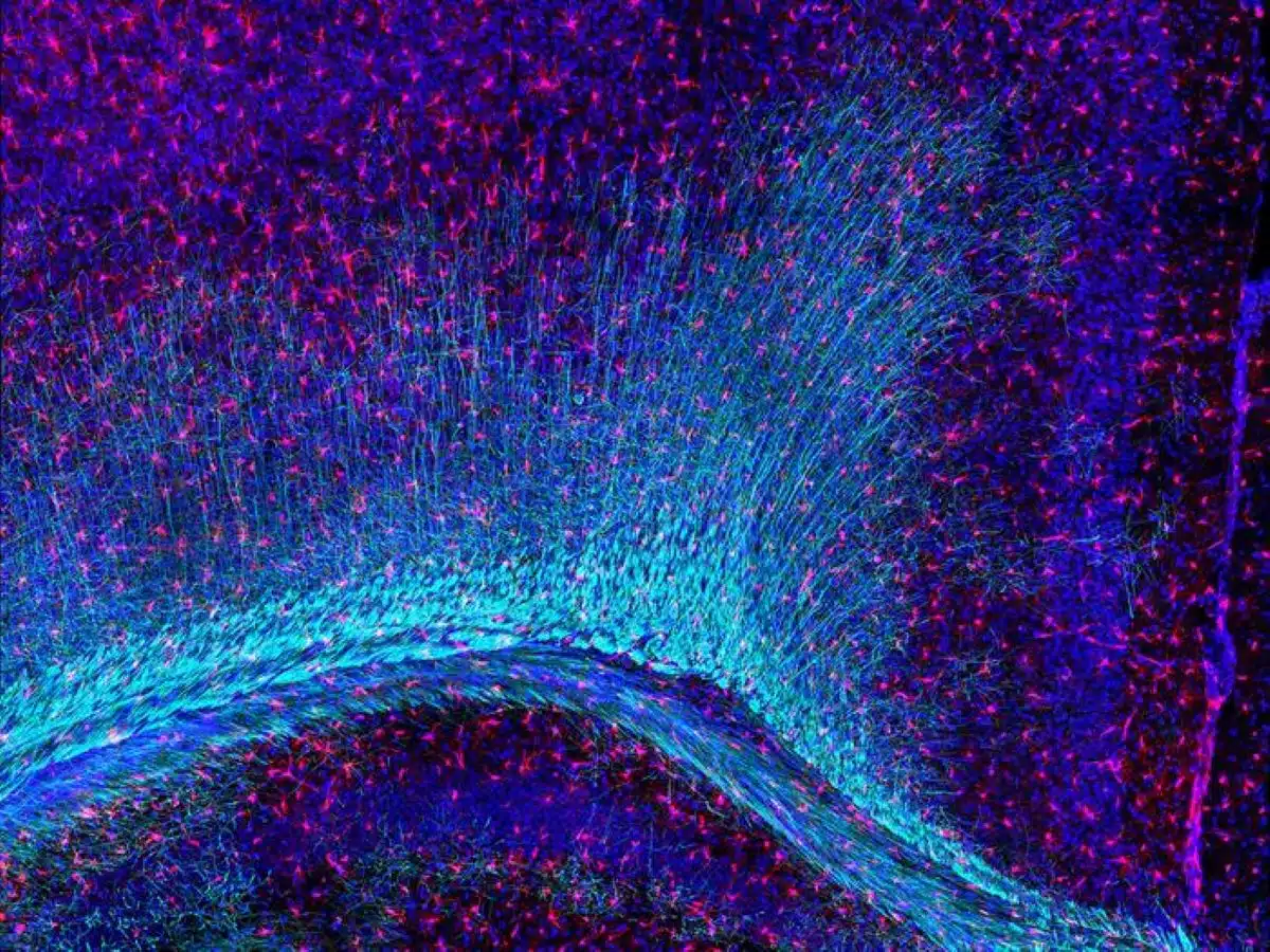
“We detected structural alterations in specific brain regions at early stages of development, as well as abnormalities in myelin, the protective sheath surrounding nerve fibers. We also identified molecular changes suggesting dysfunction in oligodendrocytes, the cells responsible for myelin production,” explains Anne-Claire Compagnion, postdoctoral researcher at the Department of Biomedical Sciences and first author of the study.
Furthermore, the study showed that microglia lacking TDP-43 lose their ability to efficiently resolve myelin abnormalities, which are naturally generated during the myelination process that occurs throughout brain development. This impairment is associated with dysfunction of a major cellular pathway known as the TREM2–DAP12 axis, which is essential for normal microglial function.
“Our findings reveal a previously unrecognized role for TDP-43 in regulating microglial function and demonstrate how its disruption could contribute to the development of neurological diseases”, concludes Rosa Chiara Paolicelli.
Key Questions Answered:
A: This discovery highlights the critical importance of cellular cross-talk in the brain. While oligodendrocytes are the specialized factory cells that build the myelin insulation, the creation process is inherently messy, naturally generating minor architectural errors and debris during childhood development. Microglia act as the clean-up crew; their job is to sweep away these structural errors to allow for clean insulation. If the microglial clean-up crew goes on strike due to a loss of TDP-43, the accumulated debris suffocates the local environment, throwing the oligodendrocyte factories into complete dysfunction.
A: Think of the TREM2–DAP12 axis as the specialized sensory array and communication link that microglia use to navigate their environment. TREM2 acts as an external sensor on the cell’s surface, listening for chemical signals that indicate cell damage or stray debris. When it spots a target, it transmits a message through its partner molecule, DAP12, telling the cell to consume and destroy the garbage. The Lausanne study proved that without TDP-43, this entire tracking network is disabled, leaving the microglia blind to the mounting myelin errors around them.
A: For decades, neurodegenerative conditions like ALS have been viewed almost exclusively as “neuron-only” problems, focusing entirely on how toxic TDP-43 clumps destroy motor nerve lines. Dr. Rosa Chiara Paolicelli’s breakthrough changes that narrative completely by proving that TDP-43 plays an equally vital, non-neuronal role inside our immune defense network. By demonstrating that losing this single protein inside microglia ruins the brain’s structural development and causes permanent movement problems, this research opens up a whole new therapeutic frontier: designing treatments to preserve immune cell housekeeping before structural decay takes hold.
Editorial Notes:
- This article was edited by a Neuroscience News editor.
- Journal paper reviewed in full.
- Additional context added by our staff.
About this psychology and diet research news
Author: Géraldine Falbriard
Source: Stellate Communications
Contact: Géraldine Falbriard – Stellate Communications
Image: The image is credited to Paolicelli Lab, Unil
Original Research: Open access.
“Microglial TDP-43 mediates myelin refinement and represses Tyrobp cryptic exon inclusion in mice” by Anne-Claire Compagnion, Andranik Ivanov, Anil Rana, Felipe Espinoza, Thomas Sandmann, Fanny S. Martineau, Katia Monsorno, Roberta Facchinetti, Alessandro Matera, Lionel Rougé, Fernando González Ibáñez, Clarissa Catale, Matteo Bizzotto, Sonia Garel, Michela Matteoli, Yutaro Kashiwagi, Ryuta Koyama, Christian Haass, Marie-Eve Tremblay, Dieter Beule, Ileana Jelescu, Valerio Zerbi, Gilbert Di Paolo & Rosa C. Paolicelli. Nature Neuroscience
DOI:10.1038/s41593-026-02348-3
Abstract
Microglial TDP-43 mediates myelin refinement and represses Tyrobp cryptic exon inclusion in mice
TDP-43 proteinopathy is a hallmark of neurodegenerative disorders such as amyotrophic lateral sclerosis and frontotemporal dementia where mislocalization of TDP-43 has been observed in neurons and glial cells.
However, the role of TDP-43 in microglia and the consequences of its loss of function remain unexplored. Combining magnetic resonance imaging, and confocal, and electron microscopy, we uncovered structural changes and myelin abnormalities in the early postnatal brain of mice lacking microglial TDP-43.
Spatial transcriptomics further revealed an enriched interferon-responsive signature associated with oligodendrocyte dysfunction. Early depletion of microglial TDP-43 led to motor deficits in adult mice.
Mechanistically, knocking out TDP-43 impaired microglial ability to engulf and degrade myelin. It also led to cryptic exon inclusion in the Tyrobp mRNA, resulting in truncated DAP12 protein, thus causing defective TREM2 signaling.
Our findings reveal a role for TDP-43 in regulating the TREM2-DAP12 axis in mice, highlighting a previously unrecognized mechanism through which TDP-43 controls microglial function.


