Summary: Researchers report people who are diabetic or who smoke are more likely to have calcification in the hippocampus. The study hypothesized hippocampal calcification may contribute to cognitive deterioration and brain atrophy.
Source: RSNA.
People who smoke or have diabetes may be at increased risk of calcifications in a region of the brain crucial to memory, according to a new study published online in the journal Radiology.
Dementia is a major public health problem that affects tens of millions of people worldwide. One focus of dementia research has been the hippocampus, a brain structure important for both short- and long-term memory storage. Alzheimer’s disease, the most common type of dementia, is associated with atrophy of the hippocampus.
Researchers have hypothesized that abnormal buildups of calcium, or calcifications, in the hippocampus may be related to vascular problems that could contribute to hippocampal atrophy and subsequent cognitive deterioration. However, published research on the association between hippocampal calcification and cognitive impairment is limited.
“We know that calcifications in the hippocampus are common, especially with increasing age,” said the study’s lead author, Esther J.M. de Brouwer, M.D., a geriatrician at the University Medical Center in Utrecht, the Netherlands. “However, we did not know if calcifications in the hippocampus related to cognitive function.”
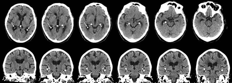
Advances in imaging have provided opportunities to explore the role of hippocampal calcifications in dementia. The development of multiplanar brain CT scans has enabled better distinction between hippocampal calcifications and calcifications in nearby brain structures like the choroid plexus.
“A multiplanar CT scan makes it possible to see the hippocampus in different anatomical planes; for example, from top to bottom, right to left and front to back,” Dr. de Brouwer said. “Before multiplanar CT scans, hippocampal calcifications were often mistaken for choroid plexus calcifications. So with multiplanar CT scans, hippocampal calcifications are better distinguished from calcifications in other areas.”
Dr. de Brouwer and colleagues studied the association between vascular risk factors like high blood pressure, diabetes and smoking, and hippocampal calcifications. They also assessed the effects of calcifications on cognitive function.
The study group included 1,991 patients, average age 78 years, who had visited a memory clinic at a Dutch hospital between 2009 and 2015. The patients had a standard diagnostic workup including cognitive tests and brain CT scans. The researchers analyzed the CT scans for the presence and severity of hippocampal calcifications.
Of the 1,991 patients, 380, or 19.1 percent, had hippocampal calcifications. Older age, diabetes and smoking were associated with an increased risk of hippocampal calcifications on CT scans.
While the study was not designed to conclusively determine if smoking and diabetes increase the risk of hippocampal calcifications, the results strongly suggest a link.
“We do think that smoking and diabetes are risk factors,” Dr. de Brouwer said. “In a recent histopathology study, hippocampal calcifications were found to be a manifestation of vascular disease. It is well known that smoking and diabetes are risk factors for cardiovascular disease. It is, therefore, likely that smoking and diabetes are risk factors for hippocampal calcifications.”
There was no link between the presence and severity of hippocampal calcifications and cognitive function; a surprising finding, according to Dr. de Brouwer, with several possible explanations.
“The hippocampus is made up of different layers, and it is possible that the calcifications did not damage the hippocampal structure that is important for memory storage,” she said. “Another explanation could be the selection of our study participants, who all came from a memory clinic.”
The researchers plan to carry out additional studies in different groups of people to better understand possible links between these calcifications and cognitive problems.
Source: Linda Brooks – RSNA
Publisher: Organized by NeuroscienceNews.com.
Image Source: NeuroscienceNews.com image is credited to RSNA.
Original Research: Open access research for “Hippocampal Calcifications: Risk Factors and Association with Cognitive Function” by Esther J. M. de Brouwer, Remko Kockelkoren, Jules J. Claus, Annemarieke de Jonghe, Mirjam I. Geerlings, Thomas E. F. Jongsma, Willem P. T. M. Mali, Jeroen Hendrikse, Pim A. de Jong, and Huiberdina L. Koek in Radiology. Published June 12 2018
doi:10.1148/radiol.2018172588
[cbtabs][cbtab title=”MLA”]RSNA “Smoking and Diabetes Linked to Brain Calcifications.” NeuroscienceNews. NeuroscienceNews, 12 June 2018.
<https://neurosciencenews.com/smoking-diabetes-brain-calcification-9316/>.[/cbtab][cbtab title=”APA”]RSNA (2018, June 12). Smoking and Diabetes Linked to Brain Calcifications. NeuroscienceNews. Retrieved June 12, 2018 from https://neurosciencenews.com/smoking-diabetes-brain-calcification-9316/[/cbtab][cbtab title=”Chicago”]RSNA “Smoking and Diabetes Linked to Brain Calcifications.” https://neurosciencenews.com/smoking-diabetes-brain-calcification-9316/ (accessed June 12, 2018).[/cbtab][/cbtabs]
Abstract
Hippocampal Calcifications: Risk Factors and Association with Cognitive Function
Purpose
To identify risk factors for hippocampal calcifications and to investigate the association between hippocampal calcifications and cognitive function.
Materials and Methods
For this retrospective study, consecutive patients visiting a memory clinic at a Dutch general hospital between April 2009 and April 2015 were identified. All individuals underwent a standard diagnostic work-up including cognitive tests and brain CT. The following vascular risk factors were assessed: hypertension, diabetes mellitus, hyperlipidemia, and smoking. Cognitive screening consisted of the Cambridge Cognitive Examination, which includes the Mini-Mental State Examination. CT scans were analyzed for the presence and severity (absent, mild, moderate, severe) of hippocampal calcifications. One measure per patient, only the most severe score, was used. Logistic regression was used to identify risk factors for hippocampal calcifications, and linear regression was used for the association between hippocampal calcifications (patient level) and cognitive function.
Results
A total of 1991 patients (mean age, 78 years; range, 45–96 years) were included. The mean age of women was 79 years (range, 47–96 years), and the mean age of men was 77 years (range, 45–95 years). Of the 1991 patients, 380 (19.1%) had hippocampal calcifications. Older age (odds ratio [OR] per year, 1.05; 95% confidence interval [CI]: 1.03, 1.06), diabetes mellitus (OR, 1.50; 95% CI: 1.12, 2.00), and smoking (OR, 1.49; 95% CI: 1.05, 2.10) were associated with the presence of hippocampal calcifications. No associations were found between presence and severity of hippocampal calcifications and cognitive function.
Conclusion
Older age, diabetes mellitus, and smoking were associated with an increased risk of hippocampal calcifications. A greater degree of hippocampal calcifications was not associated with lower cognitive function in patients with memory complaints.