Studies in mice reveal how mood-altering drugs may affect brain stem cells.
Working with mice, Johns Hopkins researchers say they have figured out how stem cells found in a part of the brain responsible for learning, memory and mood regulation decide to remain dormant or create new brain cells. Apparently, the stem cells “listen in” on the chemical communication among nearby neurons to get an idea about what is stressing the system and when they need to act.
The researchers say understanding this process of chemical signaling may shed light on how the brain reacts to its environment and how current antidepressants work, because in animals these drugs have been shown to increase the number of brain cells. The findings are reported July 29 in the advance online publication of Nature.
“What we learned is that brain stem cells don’t communicate in the official way that neurons do, through synapses or by directly signaling each other,” says Hongjun Song, Ph.D., professor of neurology and director of Johns Hopkins Medicine’s Institute for Cell Engineering’s Stem Cell Program. “Synapses, like cell phones, allow nerve cells to talk with each other. Stem cells don’t have synapses, but our experiments show they indirectly hear the neurons talking to each other; it’s like listening to someone near you talking on a phone.”
The “indirect talk” that the stem cells detect is comprised of chemical messaging fueled by the output of neurotransmitters that leak from neuronal synapses, the structures at the ends of brain cells that facilitate communication. These neurotransmitters, released from one neuron and detected by a another one, trigger receiving neurons to change their electrical charges, which either causes the neuron to fire off an electrical pulse propagating communication or to settle down, squelching further messages.

To find out which neurotransmitter brain stem cells can detect, the researchers took mouse brain tissue, attached electrodes to the stem cells and measured any change in electrical charge after the addition of certain neurotransmitters. When they treated the stem cells with the neurotransmitter GABA – a known signal-inhibiting product the stem cells’ electrical charges changed, suggesting that the stem cells can detect GABA messages.
To find out what message GABA imparts to brain stem cells, the scientists used a genetic trick to remove the gene for the GABA receptor — the protein on the surface of the cell that detects GABA — only from the brain stem cells. Microscopic observation of brain stem cells lacking the GABA receptor over five days showed these cells replicated themselves, or produced glial cells — support cells for the neurons in the brain. Brain stem cells with their GABA receptors intact appeared to stay the same, not making more cells.
Next, the team treated normal mice with valium, often used as an anti-anxiety drug and known to act like GABA by activating GABA receptors when it comes in contact with them. The scientists checked the mice on the second and seventh day of valium use and counted the number of brain stem cells in untreated mice and mice treated with the GABA activator. They found the treated mice had many more dormant stem cells than the untreated mice.
“Traditionally GABA tells neurons to shut down and not continue to propagate a message to other neurons,” says Song. “In this case the neurotransmitter also shuts off the stem cells and keeps them dormant.”
The brain stem cell population in mice (and other mammals, including humans) is surrounded by as many as 10 different kinds of intermingled neurons, says Song, and any number of these may be keeping stem cells dormant. To find out which neurons control the stem cells, the researchers inserted special light-activating proteins into the neurons that trigger the cells to send an electrical pulse, as well as to release neurotransmitter, when light shines on them. By shining light to activate a specific type of neuron and monitoring the stem cells with an electrode, Song’s team showed that one of the three types of neurons tested transmitted a signal to the stem cells causing a change in electrical charge in the stem cells. The neurons messaging the stem cells are parvalbumin-expressing interneurons.
Finally, to see if this stem cell control mechanism aligns with what an animal may be experiencing, the scientists created stress for normal mice by socially isolating them, and did the same in mice lacking GABA receptors in their brain stem cells. After a week, socially isolated normal mice had an increase in the number of stem cells and glial cells. But the socially isolated mice without GABA receptors did not show increases.
“GABA communication clearly conveys information about what brain cells experience of the outside world, and, in this case, keeps the brain stem cells in reserve, so if we don’t need them, we don’t use them up,” says Song.
Notes about this neural stem cell and neurogenesis research
Other authors on the paper include Juan Song, Chun Zhong, Michael Bonaguidi, Gerald Sun, Derek Hsu, Kimberly Christian and Guo-li Ming of Johns Hopkins University, Yan Gu and Shaoyu Ge of State University of New York at Stony Brook, Konstantinos Meletis of the Karolinska Institutet, Z. Josh Huang and Grigori Enikolopov of the Cold Spring Harbor Laboratory, Karl Deisseroth of Stanford University and Bernhard Luscher of Pennsylvania State University.
This work was supported by grants from the National Institute of Neurological Disorders and Stroke (NS047344, NS048271), the National Institute of Child Health and Human Development (HD069184), National Institute of Mental Health (MH089111), the National Institute on Aging (AG040209), the National Alliance for Research on Schizophrenia and Depression, the Adelson Medical Research Foundation, the New York State Stem Cell Science, the Ellison Medical Foundation, the Life Sciences Research Foundation and the Maryland Stem Cell Research Fund.
Contacts: Vanessa McMains & Audrey Huang – Johns Hopkins Medicine
Source: Johns Hopkins Medicine press release
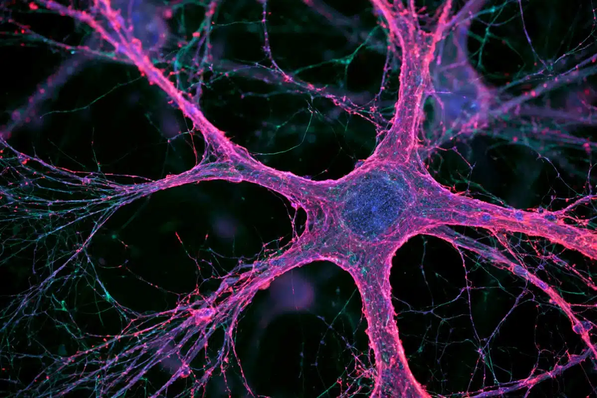
Image Source: Image adapted from Johns Hopkins Medicine press release image credited to Gerry Sun
Original research: Abstract for “Neuronal circuitry mechanism regulating adult quiescent neural stem-cell fate decision” by Juan Song, Chun Zhong, Michael A. Bonaguidi, Gerald J. Sun, Derek Hsu, Yan Gu, Konstantinos Meletis, Z. Josh Huang, Shaoyu Ge, Grigori Enikolopov, Karl Deisseroth, Bernhard Luscher, Kimberly M. Christian, Guo-li Ming & Hongjun Son in Nature 29 July 2012 doi:10.1038/nature11306