Summary: Scientists have developed human stem cell models containing the notochord, a critical tissue that guides spine and nervous system formation during embryonic development.
Using precise chemical signals inspired by natural processes in chicken, mouse, and monkey embryos, the researchers successfully coaxed human stem cells to form a notochord and a miniature trunk-like structure. This innovation mirrors key features of human development and could advance the study of spine-related birth defects and conditions like intervertebral disc degeneration.
Key Facts:
- Notochord Role: The notochord acts as a “GPS” in embryos, guiding spine and nervous system development.
- Lab-Grown Trunk: Scientists recreated notochord tissue and a trunk-like structure with organized neural and bone tissues.
- Clinical Potential: The model could aid in studying birth defects and back pain linked to intervertebral disc degeneration.
Source: Francis Crick Institute
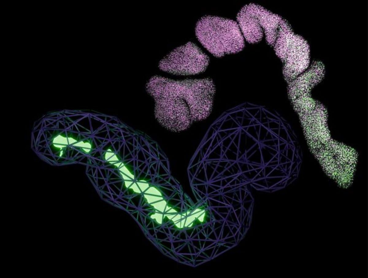
Scientists at the Francis Crick Institute have generated human stem cell model1 which, for the first time, contain notochord – a tissue in the developing embryo that acts like a navigation system, directing cells where to build the spine and nervous system (the trunk).
The work, published today in Nature, marks a significant step forward in our ability to study how the human body takes shape during early development.

The notochord, a rod-shaped tissue, is a crucial part of the scaffold of the developing body. It is a defining feature of all animals with backbones and plays a critical role in organising the tissue in the developing embryo.
Despite its importance, the complexity of the structure has meant it has been missing in previous lab-grown models of human trunk development.
In this research, the scientists first analysed chicken embryos to understand exactly how the notochord forms naturally. By comparing this with existing published information from mouse and monkey embryos, they established the timing and sequence of the molecular signals needed to create notochord tissue.
With this blueprint, they produced a precise sequence of chemical signals and used this to coax human stem cells into forming a notochord.
The stem cells formed a miniature ‘trunk-like’ structure, which spontaneously elongated to 1-2 millimetres in length. It contained developing neural tissue and bone stem cells, arranged in a pattern that mirrors development in human embryos. This suggested that the notochord was encouraging cells to become the right type of tissue at the right place at the right time.
The scientists believe this work could help to study birth defects affecting the spine and spinal cord. It could also provide insight into conditions affecting the intervertebral discs – the shock-absorbing cushions between vertebrae that develop from the notochord. These discs can cause back pain when they degenerate with age.
James Briscoe, Group Leader of the Developmental Dynamics Laboratory, and senior author of the study, said: “The notochord acts like a GPS for the developing embryo, helping to establish the body’s main axis and guiding the formation of the spine and nervous system.
“Until now, it’s been difficult to generate this vital tissue in the lab, limiting our ability to study human development and disorders. Now that we’ve created a model which works, this opens doors to study developmental conditions which we’ve been in the dark about.”
Tiago Rito, Postdoctoral Fellow in the Developmental Dynamics Laboratory, and first author of the study, said: “Finding the exact chemical signals to produce notochord was like finding the right recipe. Previous attempts to grow the notochord in the lab may have failed because we didn’t understand the required timing to add the ingredients.
“What’s particularly exciting is that the notochord in our lab-grown structures appears to function similarly to how it would in a developing embryo. It sends out chemical signals that help organise surrounding tissue, just as it would during typical development.”
About this genetics and neurodevelopment research news
Author: Clare Green
Source: Francis Crick Institute
Contact: Clare Green – Francis Crick Institute
Image: The image is credited to Tiago Rito
Original Research: Open access.
“Timely TGFβ signalling inhibition induces notochord” by Tiago Rito et al. Nature
Abstract
Timely TGFβ signalling inhibition induces notochord
The formation of the vertebrate body involves the coordinated production of trunk tissues from progenitors located in the posterior of the embryo.
Although in vitro models using pluripotent stem cells replicate aspects of this process, they lack crucial components, most notably the notochord—a defining feature of chordates that patterns surrounding tissues.
Consequently, cell types dependent on notochord signals are absent from current models of human trunk formation.
Here we performed single-cell transcriptomic analysis of chick embryos to map molecularly distinct progenitor populations and their spatial organization.
Guided by this map, we investigated how differentiating human pluripotent stem cells develop a stereotypical spatial organization of trunk cell types.
We found that YAP inactivation in conjunction with FGF-mediated MAPK signalling facilitated WNT pathway activation and induced expression of TBXT (also known as BRA). In addition, timely inhibition of WNT-induced NODAL and BMP signalling regulated the proportions of different tissue types, including notochordal cells.
This enabled us to create a three-dimensional model of human trunk development that undergoes morphogenetic movements, producing elongated structures with a notochord and ventral neural and mesodermal tissues.
Our findings provide insights into the mechanisms underlying vertebrate notochord formation and establish a more comprehensive in vitro model of human trunk development. This paves the way for future studies of tissue patterning in a physiologically relevant environment.


