

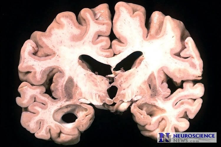
Summary: Researchers report a link between a lower BMI and greater deposits of amyloid plaques in older individuals.
Source: Mass General.
BWH/MGH study associates lower body mass index with greater deposits of Alzheimer’s-associated amyloid plaques in the brains of older individuals.
Researchers at Brigham and Women’s Hospital (BWH) and Massachusetts General Hospital (MGH) have found an association between lower weight and more extensive deposits of the Alzheimer’s-associated protein beta-amyloid in the brains of cognitively normal older individuals. The association — reported in the Journal of Alzheimer’s Disease — was seen in particular among individuals carrying the APOE4 gene variant, which is known to increase the risk of Alzheimer’s.
“Elevated cortical amyloid is believed to be the first stage of the preclinical form of Alzheimer’s disease, so our findings suggest that individuals who are underweight late in life may be at greater risk for this disease,” says Gad Marshall, MD, of the MGH and BWH Departments of Neurology, senior author of the report. “Finding this association with a strong marker of Alzheimer’s disease risk reinforces the idea that being underweight as you get older may not be a good thing when it comes to your brain health.”
While the concept of a preclinical version of Alzheimer’s disease is theoretical and not yet being used to guide clinical diagnosis or treatment, the current hypothesis involves three stages. Individuals at stage 1 are cognitively normal but have elevated amyloid deposits; stage 2 adds evidence of neurodegeneration, such as elevated tau deposits or characteristic loss of certain brain tissues, with no cognitive symptoms; and stage 3 adds cognitive changes that, while still in a normal range, indicate a decline for that individual. The current study is part of the MGH-based Harvard Aging Brain Study (HABS), designed to identify markers that predict who is likely to develop Alzheimer’s disease and how soon symptoms are likely to develop.
This investigation explored the relationship between body mass index (BMI) and beta amyloid levels in the brains of the first 280 participants to enroll in HABS, who were ages 62 to 90, cognitively normal and in good general health. Participants’ initial enrollment data included medical histories; physical exams; testing for the presence of APOE4, the major genetic risk factor for late-onset Alzheimer’s; and PET imaging with Pittsburgh compound B (PiB), which can visualize amyloid plaques in the brain.
After adjusting for factors including age, sex, education and APOE4 status, researchers found that having a lower BMI was associated with greater retention of PiB, indicating more extensive amyloid deposits in the brain. The association was most pronounced in normal-weight participants, who were the group with the lowest BMI in the study. Analysis focused on APOE status revealed that the association between lower BMI and greater PiB retention was particularly significant for individuals with the APOE4 gene variant, which is associated with increased Alzheimer’s disease risk.
Researchers hope that future studies will explain the mechanism behind the association between lower BMI and increased amyloid levels. “A likely explanation for the association is that low BMI is an indicator for frailty – a syndrome involving reduced weight, slower movement and loss of strength that is known to be associated with Alzheimer’s risk,” says Marshall, who is an assistant professor of Neurology at Harvard Medical School. “One way to get closer to determining any cause and effect relationship will be following these individuals over time to see whether their baseline BMI does predict the development of symptoms, which we are doing in HABS, and eventually investigating whether maintaining or even increasing BMI in late life has an effect on outcomes. Right now, we’re also studying whether BMI is associated with any other clinical and imaging markers of Alzheimer’s disease.”
David C. Hsu, MD, formerly with the MGH Department of Psychiatry and the BWH Department of Neurology and now at Mercy Medical Group in Sacramento, Calif., is lead author of the Journal of Alzheimer’s Disease paper.
Funding: This study was supported by the Harvard Aging Brain Study (NIH/NIA P01 AG036694, R01 AG037497, and R01 AG046396), K23 AG033634, K24 AG035007, and the Massachusetts Alzheimer’s Disease Research Center (P50AG005134).
Source: Terri Ogan – Mass General
Image Source: This NeuroscienceNews.com image is in the public domain.
Original Research: Abstract for “Lower Late-Life Body-Mass Index is Associated with Higher Cortical Amyloid Burden in Clinically Normal Elderly” by Hsu, David C.; Mormino, Elizabeth C.; Schultz, Aaron P.; Amariglio, Rebecca E.; Donovan, Nancy J.; Rentz, Dorene M.; Johnson, Keith A.; Sperling, Reisa A.; and Marshall, Gad A. in Journal of Alzheimer’s Disease. Published online June 18 2016 doi:10.3233/JAD-150987
[cbtabs][cbtab title=”MLA”]Mass General. “Lower Weight in Late Life May Increase Alzheimer’s Risk.” NeuroscienceNews. NeuroscienceNews, 2 August 2016.
<https://neurosciencenews.com/aging-weight-alzheimers-4770/>.[/cbtab][cbtab title=”APA”]Mass General. (2016, August 2). Lower Weight in Late Life May Increase Alzheimer’s Risk. NeuroscienceNews. Retrieved August 2, 2016 from https://neurosciencenews.com/aging-weight-alzheimers-4770/[/cbtab][cbtab title=”Chicago”]Mass General. “Lower Weight in Late Life May Increase Alzheimer’s Risk.” https://neurosciencenews.com/aging-weight-alzheimers-4770/ (accessed August 2, 2016).[/cbtab][/cbtabs]
Abstract
Lower Late-Life Body-Mass Index is Associated with Higher Cortical Amyloid Burden in Clinically Normal Elderly
Background: Lower body-mass index (BMI) in late life has been associated with an increased risk of dementia, and weight loss has been associated with more rapid decline in Alzheimer’s disease (AD) dementia. Objective: To explore the association between BMI and cortical amyloid burden in clinically normal (CN) elderly at risk for AD dementia.
Methods: Cross-sectional analyses were completed using baseline data from the Harvard Aging Brain Study, consisting of 280 community-dwelling CN older adults aged 62–90. Assessments included medical histories and physical exam, Pittsburgh compound B (PiB) positron emission tomography (PET) amyloid imaging, and apolipoprotein E ɛ4 (APOE4) genotyping. For the primary analysis, a general linear regression model was used to evaluate the association of BMI with PiB retention. Covariates included age, sex, years of education, and APOE4 carrier status. Secondary analyses were performed for BMI subdivisions (normal, overweight, obese), APOE4 carriers, and BMI×APOE4 interaction.
Results: In the primary analysis, greater PiB retention was associated with lower BMI (β = –0.14, p = 0.02). In the secondary analyses, APOE4 carrier status (β= –0.27, p = 0.02) and normal BMI (β= –0.25, p = 0.01), as opposed to overweight or obese BMI, were associated with greater PiB retention. The BMI×APOE4 interaction was also significant (β= –0.14, p = 0.04).
Conclusions: This finding offers new insight into the role of BMI at the preclinical stage of AD, wherein lower BMI late in life is associated with greater cortical amyloid burden. Future studies are needed to elucidate the mechanism behind this association, especially in those with lower BMI who are APOE4 carriers.
“Lower Late-Life Body-Mass Index is Associated with Higher Cortical Amyloid Burden in Clinically Normal Elderly” by Hsu, David C.; Mormino, Elizabeth C.; Schultz, Aaron P.; Amariglio, Rebecca E.; Donovan, Nancy J.; Rentz, Dorene M.; Johnson, Keith A.; Sperling, Reisa A.; and Marshall, Gad A. in Journal of Alzheimer’s Disease. Published online June 18 2016 doi:10.3233/JAD-150987


