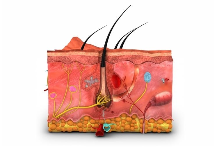
Summary: Obese mice treated with the TSLP cytokine showed a significant loss in abdominal fat and weight. The fat loss was not associated with reduced food intake or faster metabolism, instead the cytokine stimulated the immune system to release lipids via the skin’s oil-producing sebaceous glands.
Source: University of Pennsylvania
Treating obese mice with the cytokine known as TSLP led to significant abdominal fat and weight loss compared to controls, according to new research published Thursday in Science from researchers in the Perelman School of Medicine at the University of Pennsylvania.
Unexpectedly, the fat loss was not associated with decreased food intake or faster metabolism. Instead, the researchers discovered that TSLP stimulated the immune system to release lipids through the skin’s oil-producing sebaceous glands.
“This was a completely unforeseen finding, but we’ve demonstrated that fat loss can be achieved by secreting calories from the skin in the form of energy-rich sebum,” said principal investigator Taku Kambayashi, MD, PhD, an associate professor of Pathology and Laboratory Medicine at Penn, who led the study with fourth-year medical student Ruth Choa, PhD. “We believe that we are the first group to show a non-hormonal way to induce this process, highlighting an unexpected role for the body’s immune system.”
The animal model findings, Kambayashi said, support the possibility that increasing sebum production via the immune system could be a strategy for treating obesity in people.
The Hypothesis
Thymic stromal lymphopoietin (TSLP) is a cytokine — a type of immune system protein — involved in asthma and other allergic diseases. The Kambayashi research group has been investigating the expanded role of this cytokine to activate Type 2 immune cells and expand T regulatory cells. Since past studies have indicated that these cells can regulate energy metabolism, the researchers predicted that treating overweight mice with TSLP could stimulate an immune response, which could subsequently counteract some of the harmful effects of obesity.
“Initially, we did not think TSLP would have any effect on obesity itself. What we wanted to find out was whether it could impact insulin resistance,” Kambayashi said. “We thought that the cytokine could correct Type 2 diabetes, without actually causing the mice to lose any weight.”
The Experiment
To test the effect of TSLP on Type 2 diabetes, the researchers injected obese mice with a viral vector that would increase their bodies’ TSLP levels. After four weeks, the research team found that TSLP had not only affected their diabetes risk, but it had actually reversed the obesity in the mice, which were fed a high-fat diet. While the control group continued to gain weight, the weight of the TSLP-treated mice went from 45 grams down to a healthy 25 grams, on average, in just 28 days.
Most strikingly, the TSLP-treated mice also decreased their visceral fat mass. Visceral fat is the white fat that is stored in the abdomen around major organs, which can increase diabetes, heart disease, and stroke risk. These mice also experienced improved blood glucose and fasting insulin levels, as well as decreased risk of fatty liver disease.

Given the dramatic results, Kambayashi assumed that the TSLP was sickening the mice and reducing their appetites. However, after further testing, his group found that the TSLP-treated mice were actually eating 20 to 30 percent more, had similar energy expenditures, base metabolic rates, and activity levels, when compared to their non-treated counterparts.
The Findings
To explain the weight loss, Kambayashi recalled a small observation he had previously ignored: “When I looked at the coats of the TSLP-treated mice, I noticed that they glistened in the light. I always knew exactly which mice had been treated, because they were so much shinier than the others,” he said.
Kambayashi considered a far-fetched idea — was their greasy hair a sign that the mice were “sweating” out fat from their skin?
To test the theory, the researchers shaved the TSLP-treated mice and the controls and then extracted oils from their fur. They found that Kambayashi’s hypothesis was correct: The shiny fur contained sebum-specific lipids. Sebum is a calorically-dense substance produced by sebocytes (highly specialized epithelial cells) in the sebaceous glands and helps to form the skin barrier. This confirmed that the release of oil through the skin was responsible for the TSLP-induced fat loss.
The Conclusions
To examine whether TSLP could potentially play a role in the control of oil secretion in humans, the researchers then examined TSLP and a panel of 18 sebaceous gland-associated genes in a publicly-available dataset. This revealed that TSLP expression is significantly and positively correlated with sebaceous gland gene expression in healthy human skin.
The study authors write that, in humans, shifting sebum release into “high gear” could feasibly lead to the “sweating of fat” and weight loss. Kambayashi’s group plans further study to test this hypothesis.
“I don’t think we naturally control our weight by regulating sebum production, but we may be able to highjack the process and increase sebum production to cause fat loss. This could lead to novel therapeutic interventions that reverse obesity and lipid disorders,” Kambayashi said.
This research was supported by grants from the National Institutes of Health (R01-HL111501, R01-10 AI121250, R01-AR070116, T32-HL07439), the Doris Duke Charitable Foundation, and the University of Pennsylvania Medical Scientist Training Program.
Penn researchers who contributed to this work include: Junichiro Tohyama, Shogo Wada, Hu Meng, Jian Hu, Mariko Okumura, Tanner F. Robertson, Ruth-Anne Langan Pai, Arben Nace, Christian Hopkins, Elizabeth A. Jacobsen, Malay Haldar, Garret A. FitzGerald, Edward M. Behrens, Andy J. Minn, Patrick Seale, George Cotsarelis, Brian Kim, John T. Seykora, Mingyao Li, and Zoltan Arany.
About this obesity research news
Source: University of Pennsylvania
Contact: Lauren Ingeno – University of Pennsylvania
Image: The image is credited to the researchers
Original Research: Closed access.
“Thymic stromal lymphopoietin induces adipose loss through sebum hypersecretion” by Ruth Choa et al. Science
Abstract
Thymic stromal lymphopoietin induces adipose loss through sebum hypersecretion
INTRODUCTION
Obesity and its associated complications are serious global concerns. Despite growing public health initiatives, obesity rates continue to rise. Thus, there is a critical need to identify pathways that affect adiposity. Recent studies indicate that the immune system can regulate adipose tissue and its metabolic function. Type 2 immune cells, such as type 2 innate lymphoid cells (ILC2s) and eosinophils, increase the metabolic rate, whereas regulatory T cells (Treg cells) promote insulin sensitivity.
RATIONALE
Thymic stromal lymphopoietin (TSLP) is an epithelial cell cytokine that is expressed at barrier sites such as the skin, lung, and gut. Because TSLP has been shown to activate type 2 immune cells and expand Treg cells, we hypothesized that TSLP could counteract obesity and its associated complications.
RESULTS
The effect of TSLP on obesity was tested by administering a Tslp-expressing adeno-associated virus serotype 8 (TSLP-AAV) to mice. Compared with mice administered control-AAV, mice given TSLP-AAV displayed selective white adipose tissue (WAT) loss, which protected against diet-induced and genetic models of obesity, insulin resistance, and nonalcoholic steatohepatitis (NASH).
Unexpectedly, TSLP-induced WAT loss was not dependent on ILC2s, eosinophils, or Treg cells. Rather, it resulted from direct activation of either CD4+ or CD8+ αβ T cell receptor (TCRαβ) T cells by TSLP in an antigen-independent manner. The adoptive transfer of T cells from the lymph nodes of TSLP-AAV–injected mice also caused WAT loss in TSLP receptor–deficient (Tslpr–/–) mice, suggesting that TSLP-stimulated T cells retain their ability to induce WAT loss.
TSLP-induced WAT loss was not associated with decreased food intake, increased fecal caloric excretion, or increased energy metabolism. Instead, the WAT loss was associated with a notable greasy hair appearance. Thin-layer chromatography analysis of extracted hair lipids from TSLP-AAV–injected mice showed that the oleaginous substance was enriched for sebum-specific lipids. Sebum is a calorically dense substance produced by sebocytes in sebaceous glands (SGs) and helps form both the physical and immune-protective skin barrier. Skin histological analysis showed that TSLP promoted sebum secretion and turnover of sebocytes. Sebum hypersecretion was responsible for TSLP-induced WAT loss because TSLP did not induce WAT loss in asebia mice, which harbor hypomorphic SGs. TSLP also induced the migration of T cells to SGs, which was required for the enhanced sebum secretion. Inhibition of T cell migration prevented TSLP-induced sebum hypersecretion and subsequent WAT loss.
At homeostasis, TSLP and T cells controlled steady-state sebum secretion. Both Tslpr–/– and T cell–deficient mice exhibited decreased sebum secretion at baseline. Many of the fatty acids within sebum have bactericidal properties, and antimicrobial peptides (AMPs) are also secreted as part of sebum for barrier protection. Accordingly, Tslpr–/– mice expressed lower levels of sebum-associated AMPs in the skin, suggesting that endogenous TSLP plays a role in skin barrier function. This TSLP-sebum axis was also applicable to humans because the expression of TSLP and sebum-associated genes were positively correlated in skin samples from healthy individuals.
CONCLUSION
Our findings support a model in which TSLP overexpression causes WAT loss by inducing skin T cell migration and increasing sebum hypersecretion. Additionally, TSLP and T cells homeostatically regulate sebum production and skin AMP expression, highlighting an unexpected role for the adaptive immune system in the maintenance of skin barrier function.


