


Breakthrough allows Einstein scientists to probe how memories form in nerve cells.
In two studies in the January 24 issue of Science, researchers at Albert Einstein College of Medicine of Yeshiva University used advanced imaging techniques to provide a window into how the brain makes memories. These insights into the molecular basis of memory were made possible by a technological tour de force never before achieved in animals: a mouse model developed at Einstein in which molecules crucial to making memories were given fluorescent “tags” so they could be observed traveling in real time in living brain cells.
Efforts to discover how neurons make memories have long confronted a major roadblock: Neurons are extremely sensitive to any kind of disruption, yet only by probing their innermost workings can scientists view the molecular processes that culminate in memories. To peer deep into neurons without harming them, Einstein researchers developed a mouse model in which they fluorescently tagged all molecules of messenger RNA (mRNA) that code for beta-actin protein – an essential structural protein found in large amounts in brain neurons and considered a key player in making memories. mRNA is a family of RNA molecules that copy DNA’s genetic information and translate it into the proteins that make life possible.
“It’s noteworthy that we were able to develop this mouse without having to use an artificial gene or other interventions that might have disrupted neurons and called our findings into question,” said Robert Singer, Ph.D., the senior author of both papers and professor and co-chair of Einstein’s department of anatomy & structural biology and co-director of the Gruss Lipper Biophotonics Center at Einstein. He also holds the Harold and Muriel Block Chair in Anatomy & Structural Biology at Einstein.
In the research described in the two Science papers, the Einstein researchers stimulated neurons from the mouse’s hippocampus, where memories are made and stored, and then watched fluorescently glowing beta-actin mRNA molecules form in the nuclei of neurons and travel within dendrites, the neuron’s branched projections. They discovered that mRNA in neurons is regulated through a novel process described as “masking” and “unmasking,” which allows beta-actin protein to be synthesized at specific times and places and in specific amounts.
“We know the beta-actin mRNA we observed in these two papers was ‘normal’ RNA, transcribed from the mouse’s naturally occurring beta-actin gene,” said Dr. Singer. “And attaching green fluorescent protein to mRNA molecules did not affect the mice, which were healthy and able to reproduce.”
Neurons come together at synapses, where slender dendritic “spines” of neurons grasp each other, much as the fingers of one hand bind those of the other. Evidence indicates that repeated neural stimulation increases the strength of synaptic connections by changing the shape of these interlocking dendrite “fingers.” Beta-actin protein appears to strengthen these synaptic connections by altering the shape of dendritic spines. Memories are thought to be encoded when stable, long-lasting synaptic connections form between neurons in contact with each other.
The first paper describes the work of Hye Yoon Park, Ph.D., a postdoctoral student in Dr. Singer’s lab at the time and now an instructor at Einstein. Her research was instrumental in developing the mice containing fluorescent beta-actin mRNA—a process that took about three years.
Dr. Park stimulated individual hippocampal neurons of the mouse and observed newly formed beta-actin mRNA molecules within 10 to 15 minutes, indicating that nerve stimulation had caused rapid transcription of the beta-actin gene. Further observations suggested that these beta-actin mRNA molecules continuously assemble and disassemble into large and small particles, respectively. These mRNA particles were seen traveling to their destinations in dendrites where beta-actin protein would be synthesized.
In the second paper, lead author and graduate student Adina Buxbaum of Dr. Singer’s lab showed that neurons may be unique among cells in how they control the synthesis of beta-actin protein.
“Having a long, attenuated structure means that neurons face a logistical problem,” said Dr. Singer. “Their beta-actin mRNA molecules must travel throughout the cell, but neurons need to control their mRNA so that it makes beta-actin protein only in certain regions at the base of dendritic spines.”
Ms. Buxbaum’s research revealed the novel mechanism by which brain neurons handle this challenge. She found that as soon as beta-actin mRNA molecules form in the nucleus of hippocampal neurons and travel out to the cytoplasm, the mRNAs are packaged into granules and so become inaccessible for making protein. She then saw that stimulating the neuron caused these granules to fall apart, so that mRNA molecules became unmasked and available for synthesizing beta-actin protein.
But that observation raised a question: How do neurons prevent these newly liberated mRNAs from making more beta-actin protein than is desirable? “Ms. Buxbaum made the remarkable observation that mRNA’s availability in neurons is a transient phenomenon,” said Dr. Singer. “She saw that after the mRNA molecules make beta-actin protein for just a few minutes, they suddenly repackage and once again become masked. In other words, the default condition for mRNA in neurons is to be packaged and inaccessible.”
These findings suggest that neurons have developed an ingenious strategy for controlling how memory-making proteins do their job. “This observation that neurons selectively activate protein synthesis and then shut it off fits perfectly with how we think memories are made,” said Dr. Singer. “Frequent stimulation of the neuron would make mRNA available in frequent, controlled bursts, causing beta-actin protein to accumulate precisely where it’s needed to strengthen the synapse.”
To gain further insight into memory’s molecular basis, the Singer lab is developing technologies for imaging neurons in the intact brains of living mice in collaboration with another Einstein faculty member in the same department, Vladislav Verkhusha, Ph.D. Since the hippocampus resides deep in the brain, they hope to develop infrared fluorescent proteins that emit light that can pass through tissue. Another possibility is a fiberoptic device that can be inserted into the brain to observe memory-making hippocampal neurons.
Notes about this neuroscience and memory research
The titles of the two Einstein papers are: “Visualization of Dynamics of Single Endogenous mRNA as Labeled in Live Mouse,” and “Single Beta-actin mRNA Detection in Neurons Reveals a Mechanism for Regulating Its Translatability.”
In addition to the authors noted above, other Einstein authors of these papers were Bin Wu, Ph.D., Young J. Yoon, Ph.D., Melissa Lopez-Jones and Xiuhua Meng. Additional contributors are Antonia Follenzi, M.D., at Università del Piemonte orientale “Amedeo Avogadro,” Vercelli, Italy; Chiso Nwokafor and Hyungsik Lim, both at Hunter College of The City University of New York, New York, NY.
The Park study was supported by NIH grants from the National Institute of Neurological Diseases and Stroke (NS083085-19) the National Institute of General Medical Sciences (GM 084364) and the National Institute of Biomedical Imaging and Bioengineering (EB13571), a National Research Service Award (GM87122), and Einstein’s Integrated Imaging Program. The Buxbaum study was supported by grant (NS083085-19) and the Weisman Family Foundation.
Contact: Deirdre Branley – Albert Einstein College of Medicine
Source: Albert Einstein College of Medicine press release
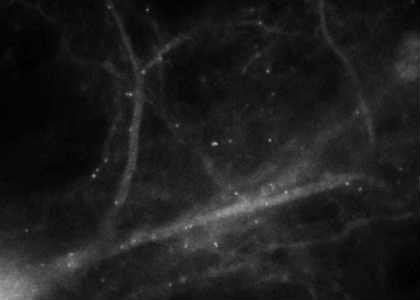
Image Source: The image is adapted from a video available at the Albert Einstein College of Medicine press release.
Original Research: Abstract for “Visualization of Dynamics of Single Endogenous mRNA Labeled in Live Mouse” by THye Yoon Park, Hyungsik Lim, Young J. Yoon, Antonia Follenzi, Chiso Nwokafor, Melissa Lopez-Jones, Xiuhua Meng, and Robert H. Singer in Science. Published online January 23 2014 doi:10.1126/science.1239200
Abstract for “Single Beta-actin mRNA Detection in Neurons Reveals a Mechanism for Regulating Its Translatability” by Adina R. Buxbaum, Bin Wu, and Robert H. Singer in Science. Published online January 23 2014 doi:10.1126/science.1242939


