

Summary: Why humans and animals must sleep remains one of the most enduring mysteries in biology. One leading framework, known as the synaptic homeostasis hypothesis, posits that the connections between neurons, synapses, gradually strengthen and multiply as we process information throughout our waking hours. This constant cellular growth consumes immense energy and clutters the brain with proteins, requiring sleep to act as a systemic “reset button” that prunes away weak connections and restores baseline balance. While this phenomenon has been thoroughly mapped in animal models, concrete human evidence has remained elusive.
Now, a pioneering study has provided direct support for this hypothesis in humans. Researchers utilized advanced positron emission tomography (PET) imaging to track a specific molecular proxy for brain cell connections in human subjects. By comparing individuals who enjoyed a full night’s rest against those subjected to 28 hours of total sleep deprivation, the team discovered measurable, widespread elevations in synaptic density markers across critical cognitive hubs of the sleep-deprived brain.
Key Facts
- The Cellular Cost of Wakefulness: Prolonged wakefulness causes the physical connections between human brain cells to strengthen and build up, demanding higher metabolic energy and validating the synaptic homeostasis model of sleep.
- SV2A as a Structural Window: Researchers used PET scans to track Synaptic Vesicle Glycoprotein 2A (SV2A), an established biomarker that directly reflects the density of active synaptic connections in the living human brain.
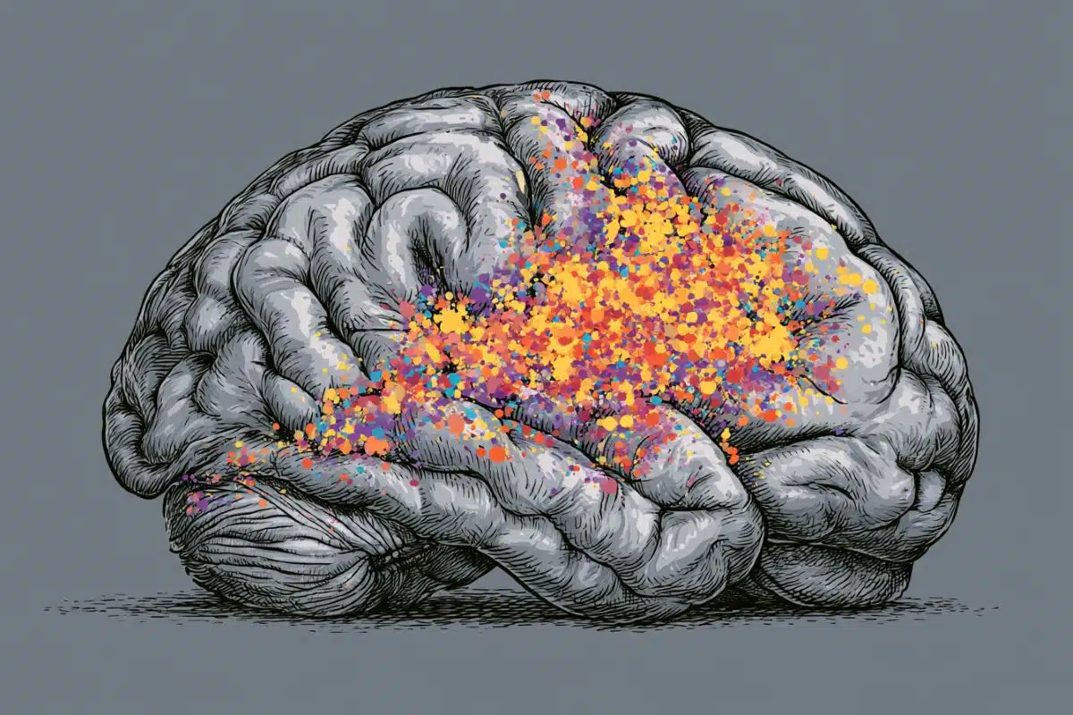
- Targeted Brain Inundation: Following 28 hours of continuous sleep deprivation, participants exhibited distinct elevations of SV2A in the hippocampus (the brain’s memory epicentre) and the thalamus (the vital sensory and information relay station).
- The Deep Sleep Push: When sleep-deprived individuals were granted a short two-hour recovery nap, those with the highest accumulated SV2A levels exhibited significantly more slow-wave activity, a direct neurological indicator of high sleep pressure and intense deep sleep.
- From Fatigue to Physical Change: The findings prove that sleep deprivation does not simply produce a psychological feeling of fatigue; it forces measurable, physical, and potentially inefficient structural shifts across human neural networks.
Source: PLOS
A night without sleep produced increased markers of connections between brain cells, showing that sleep in humans may be important for restoring cellular balance in the brain, according to a study published June 23rd in the open access journal PLOS Biology by David Elmenhorst from the Forschungszentrum Jülich Institute of Neuroscience and Medicine in North Rhine-Westphalia, Germany, and colleagues.
Scientists have long wondered why exactly humans and other animals need to sleep. One potential mechanism is that sleep is required to restore synaptic connections and homeostasis in the brain. Synapses—the connections between brain cells—become stronger during wakefulness. This increases the amount of energy the brain needs and leads to buildup of proteins in the brain.
Sleep is thought to reset these levels, reducing synaptic connections and restoring homeostasis, but evidence has thus far been limited to animal models. To determine whether the synaptic homeostasis hypothesis is supported in humans, the authors of this study used positron emission tomography (PET) to look for markers of synaptic vesicle glycoprotein 2A (SV2A), a marker of brain synapses in 40 participants, half of whom had gone one night without sleep.
The authors found that after 28 hours of continuous wakefulness, the sleep deprivation group had higher measures of SV2A in several brain regions, including the hippocampus (an area important for memory), and the thalamus, an important information relay in the brain.
When the sleep-deprived participants were allowed a two-hour nap, higher levels of SV2A were associated with more slow wave activity during sleep, a marker of deep sleep and sleep pressure. While SV2A is only a proxy for brain cell connections and the elevations were relatively small, the results support the synaptic homeostasis model of sleep, and suggest a biological connection between the need for sleep and the buildup of cell connections.
The authors add, “During sleep deprivation, the brain remains awake longer and continues to process stimuli and information. Our study shows that after approximately 28.5 hours of wakefulness, a marker for synaptic density increases in several brain regions. This suggests that sleep deprivation not only causes fatigue but is also accompanied by measurable changes in neural connections.”
Key Questions Answered:
A: SV2A stands for Synaptic Vesicle Glycoprotein 2A. It is an essential protein residing inside the microscopic sacs (vesicles) that hold chemical messengers within our brain cell connections. Because SV2A is present across virtually all synapses in the central nervous system, neuroscientists use it as a highly reliable “proxy” or molecular ruler to measure synaptic density. By tracking SV2A with PET scans, the researchers could look inside the living human skull and see whether the overall volume of active neural connections was growing or shrinking based on sleep habits.
A: For years, the Synaptic Homeostasis Hypothesis argued that wakefulness is the price the brain pays for learning. As you go about your day, your synapses must get stronger to record new information, which eventually leads to a chaotic, energy-hogging saturation of the brain. SHY claims sleep exists to systematically downscale or “turn down the volume” on these connections so the brain doesn’t short-circuit. By showing that 28 hours of wakefulness physically drives up SV2A markers in humans, this study provides the missing clinical evidence that wakefulness does indeed overload human synapses, and that sleep pressure is the body’s mechanism to force a cleanup.
A: In a living human brain, massive, sudden spikes in synaptic structural proteins would be highly dangerous and indicative of severe pathology, like a seizure. The fact that the elevations were relatively small is exactly what scientists expect from a normal, highly regulated homeostatic system. Even these minor, localized increases in the hippocampus and thalamus were strong enough to alter brain physics, directly predicting how intensely the brain fell into deep, slow-wave sleep during a nap. It shifts our understanding of sleep deprivation from a vague chemical imbalance to a tangible structural bottleneck.
Editorial Notes:
- This article was edited by a Neuroscience News editor.
- Journal paper reviewed in full.
- Additional context added by our staff.
About this sleep and neuroscience research news
Author: Claire Turner
Source: PLOS
Contact: Claire Turner – PLOS
Image: The image is credited to Neuroscience News
Original Research: Open access.
“Learning engages transient and sustained cellular mechanisms in the human brain” by Guillermina Griffa, Marco Palombo, Abraham Yeffal, Hong-Hsi Lee, Agustin Solano, Susie Y. Huang, and Valeria Della-Maggiore. PLOS Biology
DOI:10.1371/journal.pbio.3003861
Abstract
Learning engages transient and sustained cellular mechanisms in the human brain
Structural neuroplasticity supports learning, development, and shapes vulnerability to brain disorders, making it a central priority in neuroscience research. However, progress in humans has remained limited by the inability to probe cellular processes in vivo, leaving mechanistic insight largely dependent on animal models.
To address this gap, here we combined the sub-voxel sensitivity of ultra–high-gradient diffusion MRI with the cell-compartment specificity of the Soma and Neurite Density Imaging (SANDI) model to probe structural plasticity directly in the living human brain.
By tracking how learning modulates the temporal dynamics of cell bodies and cell processes, we aimed to distinguish plastic from nonplastic biological processes driving changes in microstructure. We found that learning a motor skill triggered two distinct temporal responses: a transient expansion of cell bodies across all brain regions engaged by the task, consistent with a short-lived homeostatic mechanism, and a sustained increase in cell-process density restricted to key motor regions, consistent with structural plasticity.
Our approach provides a mechanistic window into human neuroplasticity and marks a significant step toward bridging the gap between animal and human neuroscience.


