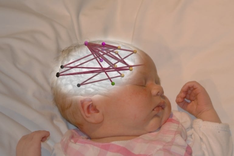
Summary: Fetal exposure to antidepressants or antiepileptic medications may affect the development of newborn brain networks.
Source: University of Helsinki
New study demonstrates that in utero exposure to mother’s antiepileptic or antidepressant medication may affect development of the newborn brain networks. In the study, novel mathematical methods were developed to allow future research on how commonly used drugs or other environmental conditions affect the newborn brain.
Pregnant mothers may need treatment for their medical conditions, such as mood disorder or epilepsy.
The effects of such drug treatment on newborn brain network functions was examined in a study conducted at the BABA Center, a research unit in University of Helsinki and New Children’s Hospital of HUS Helsinki University Hospital.
The study used electroencephalography (EEG) to measure electrical brain activity during sleep, and cortical network properties were calculated using advanced mathematical techniques.
“In prior studies, we have shown that changes in cortical activity across sleep states may provide important information on infants’ neurological condition,” Senior Researcher Anton Tokariev says.
The study demonstrated that exposure to antiepileptics and antidepressants during the fetal period leads to widespread changes in the cortical networks, and these effects may be specific to the type of drug exposure.
In the case of antidepressants, the effect was more pronounced in local cortical networks. In contrast, exposure to antiepileptics had drug-specific effects on brain wide networks. Both drug types affected brain networks that are reactive to changes in sleep stages.
“What was clinically significant in the findings was that, some EEG findings linked to children’s subsequent neuropsychological development. Stronger changes in neural networks predicted a greater deviation in development at two years of age,” says Mari Videman, specialist in paediatric neurology at HUS Helsinki University Hospital.
Shedding new light on early brain development
The studies offer an entirely new way of assessing the effects of pharmaceutical agents on the development of child’s brain function.
“The EEG measurement technique developed at the BABA Center and its associated state-of-the-art mathematical assessment of the brain’s neural networks constitute breakthroughs in clinical research on early neurodevelopment,” Professor Sampsa Vanhatalo says.
Vanhatalo considers it particularly important that these EEG -based measures open a window into mechanisms that operate between neuronal cell. This leads to an opportunitity to compare results observed in human children with research conducted using laboratory-animal models.

Such translational work is needed to understand the mechanistic underpinnings of the drug effects. For instance, identical animal work is required to study how the amount or timing of maternal drug treatment would affect brain function of the offspring.
“Our novel methods provide a general analytical framework to support extensive future research on the questions how fetal brain development is affected by changes in intrauterine environment. Such studies may go far beyond maternal drug treatment, including also mother’s nutrition and overall physical condition, as well as myriad of further environmental factors,” Vanhatalo summarises.
About this neurodevelopment and pharmacology research news
Author: Anton Tokariev
Source: University of Helsinki
Contact: Anton Tokariev – University of Helsinki
Image: The image is credited to Sampsa Vanhatalo
Original Research: Open access.
“Cortical Cross-Frequency Coupling Is Affected by in utero Exposure to Antidepressant Medication” by Anton Tokariev et al. Frontiers in Neuroscience
Abstract
Cortical Cross-Frequency Coupling Is Affected by in utero Exposure to Antidepressant Medication
Up to five percent of human infants are exposed to maternal antidepressant medication by serotonin reuptake inhibitors (SRI) during pregnancy, yet the SRI effects on infants’ early neurodevelopment are not fully understood.
Here, we studied how maternal SRI medication affects cortical frequency-specific and cross-frequency interactions estimated, respectively, by phase-phase correlations (PPC) and phase-amplitude coupling (PAC) in electroencephalographic (EEG) recordings.
We examined the cortical activity in infants after fetal exposure to SRIs relative to a control group of infants without medical history of any kind.
Our findings show that the sleep-related dynamics of PPC networks are selectively affected by in utero SRI exposure, however, those alterations do not correlate to later neurocognitive development as tested by neuropsychological evaluation at two years of age.
In turn, phase-amplitude coupling was found to be suppressed in SRI infants across multiple distributed cortical regions and these effects were linked to their neurocognitive outcomes.
Our results are compatible with the overall notion that in utero drug exposures may cause subtle, yet measurable changes in the brain structure and function.
Our present findings are based on the measures of local and inter-areal neuronal interactions in the cortex which can be readily used across species, as well as between different scales of inspection: from the whole animals to in vitro preparations.
Therefore, this work opens a framework to explore the cellular and molecular mechanisms underlying neurodevelopmental SRI effects at all translational levels.


