

fMRI identifies differences in response of mothers’ brains to images of their child and their dog.
It has become common for people who have pets to refer to themselves as “pet parents,” but how closely does the relationship between people and their non-human companions mirror the parent-child relationship? A small study from a group of Massachusetts General Hospital (MGH) researchers makes a contribution to answering this complex question by investigating differences in how important brain structures are activated when women view images of their children and of their own dogs. Their report is being published in the open-access journal PLOS ONE.
“Pets hold a special place in many people’s hearts and lives, and there is compelling evidence from clinical and laboratory studies that interacting with pets can be beneficial to the physical, social and emotional wellbeing of humans,” says Lori Palley, DVM, of the MGH Center for Comparative Medicine, co-lead author of the report. “Several previous studies have found that levels of neurohormones like oxytocin – which is involved in pair-bonding and maternal attachment – rise after interaction with pets, and new brain imaging technologies are helping us begin to understand the neurobiological basis of the relationship, which is exciting.”
In order to compare patterns of brain activation involved with the human-pet bond with those elicited by the maternal-child bond, the study enrolled a group of women with at least one child aged 2 to 10 years old and one pet dog that had been in the household for two years or longer. Participation consisted of two sessions, the first being a home visit during which participants completed several questionnaires, including ones regarding their relationships with both their child and pet dog. The participants’ dog and child were also photographed in each participants’ home.
The second session took place at the Athinoula A. Martinos Center for Biomedical Imaging at MGH, where functional magnetic resonance imaging (fMRI) – which indicates levels of activation in specific brain structures by detecting changes in blood flow and oxygen levels – was performed as participants lay in a scanner and viewed a series of photographs. The photos included images of each participant’s own child and own dog alternating with those of an unfamiliar child and dog belonging to another study participant. After the scanning session, each participant completed additional assessments, including an image recognition test to confirm she had paid close attention to photos presented during scanning, and rated several images from each category shown during the session on factors relating to pleasantness and excitement.
Of 16 women originally enrolled, complete information and MR data was available for 14 participants. The imaging studies revealed both similarities and differences in the way important brain regions reacted to images of a woman’s own child and own dog. Areas previously reported as important for functions such as emotion, reward, affiliation, visual processing and social interaction all showed increased activity when participants viewed either their own child or their own dog. A region known to be important to bond formation – the substantia nigra/ventral tegmental area (SNi/VTA) – was activated only in response to images of a participant’s own child. The fusiform gyrus, which is involved in facial recognition and other visual processing functions, actually showed greater response to own-dog images than own-child images.
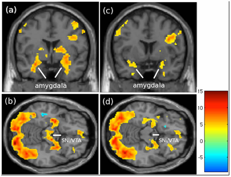
“Although this is a small study that may not apply to other individuals, the results suggest there is a common brain network important for pair-bond formation and maintenance that is activated when mothers viewed images of either their child or their dog,” says Luke Stoeckel, PhD, MGH Department of Psychiatry, co-lead author of the PLOS One report. “We also observed differences in activation of some regions that may reflect variance in the evolutionary course and function of these relationships. For example, like the SNi/VTA, the nucleus accumbens has been reported to have an important role in pair-bonding in both human and animal studies. But that region showed greater deactivation when mothers viewed their own-dog images instead of greater activation in response to own-child images, as one might expect. We think the greater response of the fusiform gyrus to images of participants’ dogs may reflect the increased reliance on visual than verbal cues in human-animal communications.”
Co-author Randy Gollub, MD, PhD, of MGH Psychiatry adds, “Since fMRI is an indirect measure of neural activity and can only correlate brain activity with an individual’s experience, it will be interesting to see if future studies can directly test whether these patterns of brain activity are explained by the specific cognitive and emotional functions involved in human-animal relationships. Further, the similarities and differences in brain activity revealed by functional neuroimaging may help to generate hypotheses that eventually provide an explanation for the complexities underlying human-animal relationships.”
The investigators note that further research is needed to replicate these findings in a larger sample and to see if they are seen in other populations – such as women without children, fathers and parents of adopted children – and in relationships with other animal species. Combining fMRI studies with additional behavioral and physiological measures could obtain evidence to support a direct relationship between the observed brain activity and the purported functions.
Stoeckel is a clinical neuropsychologist and lecturer on psychology, and Gollub an associate professor of Psychiatry at Harvard Medical School. Additional co-authors of the PLOS ONE report are Eden Evins, MD, MGH Psychiatry, and Steven Niemi, DVM, Harvard University. Support for the study includes National Institutes of Health grants K23DA032612 and K24DA030443 and support from the Charles A. King Trust. The study was facilitated by imaging consult support from Harvard Catalyst.
Contact: Noah Brown – Massachusetts General Hospital
Source: Massachusetts General Hospital press release
Image Source: The dog image is credited to jeremy8 / 7 and is in the public domain. The fMRI brain scans are credited to Stoeckel et al/PLOS ONE and are adapted from the open access research paper
Original Research: Full open access research for “Patterns of Brain Activation when Mothers View Their Own Child and Dog: An fMRI Study” by Luke E. Stoeckel, Lori S. Palley, Randy L. Gollub, Steven M. Niemi, and Anne Eden Evins in PLOS ONE. Published online October 3 2014 doi:10.1371/journal.pone.0107205
Patterns of Brain Activation when Mothers View Their Own Child and Dog: An fMRI Study
Neural substrates underlying the human-pet relationship are largely unknown. We examined fMRI brain activation patterns as mothers viewed images of their own child and dog and an unfamiliar child and dog. There was a common network of brain regions involved in emotion, reward, affiliation, visual processing and social cognition when mothers viewed images of both their child and dog. Viewing images of their child resulted in brain activity in the midbrain (ventral tegmental area/substantia nigra involved in reward/affiliation), while a more posterior cortical brain activation pattern involving fusiform gyrus (visual processing of faces and social cognition) characterized a mother’s response to her dog. Mothers also rated images of their child and dog as eliciting similar levels of excitement (arousal) and pleasantness (valence), although the difference in the own vs. unfamiliar child comparison was larger than the own vs. unfamiliar dog comparison for arousal. Valence ratings of their dog were also positively correlated with ratings of the attachment to their dog. Although there are similarities in the perceived emotional experience and brain function associated with the mother-child and mother-dog bond, there are also key differences that may reflect variance in the evolutionary course and function of these relationships.
“Patterns of Brain Activation when Mothers View Their Own Child and Dog: An fMRI Study” by Luke E. Stoeckel, Lori S. Palley, Randy L. Gollub, Steven M. Niemi, and Anne Eden Evins in Development and Psychopathology, October 3 2014 doi:10.1371/journal.pone.0107205.


