

Summary: A new study reveals how microglia help clear dead and damaged materials away from healthy, neighboring neurons following injury.
Source: Rockefeller University Press.
Researchers at the University of Virginia School of Medicine have discovered that microglia, specialized immune cells in the brain, play a key role in clearing dead material after brain injury. The study, which will be published June 25 in the Journal of Experimental Medicine, reveals that microglia gobble up the remnants of injured neurons, which could prevent the damage from spreading to neighboring neurons and causing more extensive neurodegeneration.
In every tissue of the body, dead and dying cells must quickly be removed to prevent the development of inflammation, which could trigger the death of neighboring cells. This removal is carried out by specialized cells that engulf and break down cellular debris, otherwise known as phagocytic cells. But scientists have yet to determine which cells are responsible for removing neuronal debris when the brain or spinal cord is damaged.
Jonathan Kipnis, chairman of UVA’s Department of Neuroscience, and his colleagues examined injuries to the optic nerve of mice, which cause retinal ganglion neurons to degenerate and leave debris in a distant region of the brain. The researchers found that this debris is engulfed by microglia.
Microglia, which permanently reside in the central nervous system, are a type of phagocytic cell that can engulf bacteria and other pathogens that have infected the brain. They also play an important role in the developing brain, pruning away neuronal synapses that have failed to become fully active.
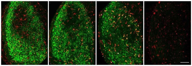
In adult brains, microglia appear to recognize degenerating neurons using some of the same molecules they use to recognize inactive synapses or invading pathogens. Kipnis and colleagues found that, after optic nerve injury, microglia produce “complement” proteins that help the phagocytic cells identify their targets.
The researchers studied what happened after optic nerve injury in mice when microglia did not produce “complement” proteins and found that the microglia did not clear the debris.
“In the future, we hope to further identify how microglia are activated in response to neurodegeneration and how they then remove neuronal debris,” says Kipnis, director of UVA’s Center for Brain Immunology and Glia (BIG). “Knowing these mechanisms might allow us to boost the clearance of potentially toxic debris by microglia and limit the spread of neurodegeneration following brain or spinal cord injury.”
Funding: NIH funded study.
Source: Rory Williams – Rockefeller University Press
Publisher: Organized by NeuroscienceNews.com.
Image Source: NeuroscienceNews.com image is credited to Norris et al., 2018.
Original Research: Open access research for “Neuronal integrity and complement control synaptic material clearance by microglia after CNS injury” by Geoffrey T. Norris, Igor Smirnov, Anthony J. Filiano, Hannah M. Shadowen, Kris R. Cody, Jeremy A. Thompson, Tajie H. Harris, Alban Gaultier, Christopher C. Overall, and Jonathan Kipnis in Journal of Experimental Medicine. Published June 25 2018.
doi:10.1084/jem.20172244
[cbtabs][cbtab title=”MLA”]Rockefeller University Press “Researchers Identify Brain Cells Responsible for Removing Damaged Neurons After Injury .” NeuroscienceNews. NeuroscienceNews, 25 June 2018.
<https://neurosciencenews.com/microglia-neuron-damage-9439 />.[/cbtab][cbtab title=”APA”]Rockefeller University Press (2018, June 25). Researchers Identify Brain Cells Responsible for Removing Damaged Neurons After Injury . NeuroscienceNews. Retrieved June 25, 2018 from https://neurosciencenews.com/microglia-neuron-damage-9439 /[/cbtab][cbtab title=”Chicago”]Rockefeller University Press “Researchers Identify Brain Cells Responsible for Removing Damaged Neurons After Injury .” https://neurosciencenews.com/microglia-neuron-damage-9439 / (accessed June 25, 2018).[/cbtab][/cbtabs]
Abstract
Neuronal integrity and complement control synaptic material clearance by microglia after CNS injury
Phagocytosis of synaptic material by microglia is critical for central nervous system development. Less well understood is this microglial function in the injured adult brain. Assay of microglial phagocytosis is challenging, because peripheral myeloid cells engraft the site of injury, which could obscure interpretation of microglial roles. The model used here, optic nerve crush injury, results in degeneration of synapses in the dorsal lateral geniculate nucleus (dLGN), which stimulates rapid activation and engulfment of synaptic material by resident microglia without myeloid cell engraftment. Pharmacological depletion of microglia causes postinjury accumulation of synaptic debris, suggesting that microglia are the dominant postinjury phagocytes. Genetic or pharmacological manipulations revealed that neuronal activity does not trigger microglia phagocytosis after injury. RNA sequencing reveals C1q and CD11b/CR3 involvement in clearance of debris by dLGN-resident microglia. Indeed, C1qa−/− and Itgam−/− mice exhibit impaired postinjury debris clearance. Our results show how neurodegenerative debris is cleared by microglia and offers a model for studying its mechanisms and physiological roles.


