

Summary: Using optogenetics to inhibit the JNK protein prevented synapses from shrinking in response to stress.
Source: University of Turku
New study from Eleanor Coffey’s lab at Turku Bioscience Centre in Finland identifies that the JNK protein triggers nerve cells to withdraw their synapses when stressed.
Synapses are tiny cell protrusions where electrochemical impulses pass between nerves. Prolonged stress in the brain causes synapse withdrawal and maladaptive changes to circuits that are linked to the development of major depressive disorder.
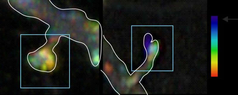
Postdoctoral Researcher Patrik Hollós and colleagues used a light-activated optogenetic tool to switch off the activity of a protein called JNK specifically in synapses.
“Using a light beam to inhibit the JNK protein prevented synapses from shrinking in response to stress. Specifically the internalisation of a receptor called “AMPAR”, an early event in synapse disassembly, was blocked,” explains Hollós.
JNK Is a Stress Sensor in Synapses and May Elicit the Effects of Ketamine
Researchers also found that the novel, fast-acting anti-depressant ketamine inhibited the JNK protein while preventing synapse retraction.
“These results show that the JNK protein is a stress sensor in synapses. When activated, it triggers the disassembly of synapse machinery followed by rapid synapse regression. Conversely, inhibiting the JNK protein makes synapses able to withstand chronic endocrine stress. This may be relevant for conditions where hormonal stress leads to synapse elimination but also to control synapse number under normal homeostatic conditions,” says team leader Eleanor Coffey.
These findings help us to understand how stress dismantles synapses, and provides clues for novel targeted therapies.
Research was conducted at Turku Bioscience Centre a joint research facility of the University of Turku and Åbo Akademi University.
Source:
University of Turku
Media Contacts:
Eleanor Coffey – University of Turku
Image Source:
The image is credited to Turku Bioscience.
Original Research: Open access
“Optogenetic Control of Spine-Head JNK Reveals a Role in Dendritic Spine Regression”. Patrik Hollos, Jismi M. John, Jukka V. Lehtonen and Eleanor T. Coffey.
eNeuro doi:10.1523/ENEURO.0303-19.2019.
Abstract
Optogenetic Control of Spine-Head JNK Reveals a Role in Dendritic Spine Regression
In this study, we use an optogenetic inhibitor of c-Jun NH2-terminal kinase (JNK) in dendritic spine sub-compartments of rat hippocampal neurons. We show that JNK inhibition exerts rapid (within seconds) reorganization of actin in the spine-head. Using real-time Förster resonance energy transfer (FRET) to measure JNK activity, we find that either excitotoxic insult (NMDA) or endocrine stress (corticosterone), activate spine-head JNK causing internalization of AMPARs and spine retraction. Both events are prevented upon optogenetic inhibition of JNK, and rescued by JNK inhibition even 2 h after insult. Moreover, we identify that the fast-acting anti-depressant ketamine reduces JNK activity in hippocampal neurons suggesting that JNK inhibition may be a downstream mediator of its anti-depressant effect. In conclusion, we show that JNK activation plays a role in triggering spine elimination by NMDA or corticosterone stress, whereas inhibition of JNK facilitates regrowth of spines even in the continued presence of glucocorticoid. This identifies that JNK acts locally in the spine-head to promote AMPAR internalization and spine shrinkage following stress, and reveals a protective function for JNK inhibition in preventing spine regression.


