

New findings by Columbia researchers suggest that along with amyloid deposits, white matter hyperintensities (WMHs) may be a second necessary factor for the development of Alzheimer’s disease.
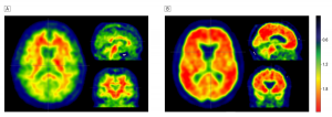
Most current approaches to Alzheimer’s disease focus on the accumulation of amyloid plaque in the brain. The researchers at the Taub Institute for Research on Alzheimer’s Disease and the Aging Brain, led by Adam M. Brickman, PhD, assistant professor of neuropsychology, examined the additional contribution of small-vessel cerebrovascular disease, which they visualized as white matter hyperintensities (WMHs).
The study included 20 subjects with clinically defined Alzheimer’s disease, 59 subjects with mild cognitive impairment, and 21 normal control subjects. Using data from the Alzheimer’s Disease Neuroimaging Initiative public database, the researchers found that amyloid and WHMs were equally associated with an Alzheimer’s diagnosis. Amyloid and WMHs were also equally predictive of which subjects with mildcognitive impairment would go on to develop Alzheimer’s. Among those with significant amyloid, WMHs were more prevalent in those with Alzheimer’s than in normal control subjects.
Because the risk factors for WMHs, which are mainly vascular, can be controlled, the findings suggest potential ways to prevent the development of Alzheimer’s in those with amyloid deposits.
The research, “White Matter Hyperintensities and Cerebral Amyloidosis” is published in JAMA Neurology.
Notes about this Alzheimer’s disease research
The other authors are Frank A. Provenzano, MS (CUMC and Fu Foundation School of Engineering and Applied Sciences); Jordan Muraskin, MS (CUMC and Fu Foundation School of Engineering and Applied Sciences); Guiseppe Tosto, MD (CUMC); Atul Narkhede, MS (CUMC); Ben T. Wasserman, AB (CUMC); Erica Y Griffith, BS (CUMC); Vanessa A. Guzman, BA (CUMC); Irene B. Meier, MSc (CUMC); and Molly E. Zimmerman, PhD (Albert Einstein College of Medicine, NY, NY).
The research was supported by NIH (U01 AG024904, P30 AG010129, K01 AG030514, AG029949, and AG034189).
Contact: Karin Eskenazi – Columbia University
Source: Columbia University press release
Image Source: Adapted from the Columbia University press release.
Original Research: Abstract for “White Matter Hyperintensities and Cerebral Amyloidosis” by Frank A. Provenzano, MS, Jordan Muraskin, MS, Giuseppe Tosto, MD, Atul Narkhede, MS, Ben T. Wasserman, AB, Erica Y. Griffith, BS, Vanessa A. Guzman, BA, Irene B. Meier, MSc, Molly E. Zimmerman, PhD and Adam M. Brickman, PhD in JAMA Neurology. Published online February 18 2013 doi:10.1001/jamaneurol.2013.1321


