

Summary: A new study tests conventional theories on the development of the nervous system and sheds light on how the nervous system may have evolved.
Source: Penn State.
Penn State researchers at the Huck Institutes of the Life Sciences uncover a long-standing, fundamental error in the story of the nervous system’s evolution.
A component of vertebrate neurons – known as the axon initial segment (AIS) – that is responsible for regulating the nerve cell’s output has long been thought by scientists to have evolved relatively recently, and specifically in vertebrates, in order to enable rapid, precise signaling in the complex circuitry of the vertebrate nervous system.
But a discovery made by the labs of Penn State researchers Melissa Rolls and Tim Jegla is bringing about a rewriting of a fundamental aspect of that story.
“One of the things I’ve been doing for a very long time,” said Jegla, “is studying nervous system evolution, and in general what we find is that most fundamental features of neurons – from the molecules that control the signaling to various cellular structures – tend to be old and conserved and shared between many extant species, like all the bilaterians, and the signaling molecules are even in very early nervous systems in cnidarians.”
“But one of the exceptions to this in the literature – coincidentally, the story we’re rewriting,” he said, “is that this very special area of a neuron called the axon initial segment, which is the beginning of the output end of the neuron, is a unique vertebrate adaptation for rapid signaling. The axon initial segment provides a barrier to separate the membrane of the cell body and the input end – the dendrites – from the axon, which is critical because this membrane has a different role, which is to initiate and then carry the action potential – the output of the neuron. You’ve got all these inputs coming in from the cell body and the dendrites, and it’s at the axon initial segment where the decision is made whether all those inputs warrant signaling the next cell, or not. So it’s the decision point for neurons.”
Jegla further explains that the structure and function of the axon initial segment are dependent on a protein known as ankyrin, specifically a giant isoform called ankyrin G that is known to be vertebrate-specific. This fact, coupled with results from “really old physiological studies of invertebrate neurons, structurally distinct ones that don’t even look like our neurons,” led to the widely accepted notion that the axon initial segment and the giant ankyrin isoforms, including ankyrin G, “evolved specifically in the vertebrate lineage,” he says.
But Rolls had an inkling – and data indicating – that there might be something missing from the story.
Collaborative corroboration
“I had these data generated independently by two undergraduates in my lab, showing that there is a diffusion barrier at the beginning of sensory neuron axons in fruit flies, and that there is a diffusion barrier in the axon initial segment that’s ankyrin-dependent, she said. “But we didn’t have enough data to really change this whole feeling in the field that there’s not a similar structure to the axon initial segment in fruit flies, and so those data sat around for a long time. Later we added some more pieces, but that still wasn’t quite enough to convince people that this really was the same structure.”
So Jegla began piecing together, through phylogenomic analysis, the evolutionary history of the ankyrin family.
“When I started to look at that it became clear that the accepted story of the giant isoforms of ankyrin such as ankyrin G being recently evolved and vertebrate-specific was completely wrong,” Jegla said.
The giant ankyrins, he explains, are in fact ancient forms that evolved in a bilaterian ancestor. And after mapping the genomic regions encoding these giant ankyrin isoforms across a number of metazoan and closely related lineages, Jegla determined that the largest exon was in the genome of Drosophila – Rolls’ model organism of choice, the fruit fly.
With Jegla’s new results, Rolls put this together with data her students had gathered on the function of the fruit fly’s AIS-like diffusion barrier, using a technique known as fluorescent recovery after photobleaching (FRAP).
“The way FRAP works is that if we tag a plasma membrane protein with a fluorescent molecule, we can shine an intense laser light on a region of the neuron and inactivate all the fluorophores in that region; then over time we can look to see whether other fluorophores migrate into that region, which gives us an idea about diffusion and whether these things are mobile in that particular region,” Jegla explained. “So we compared how these tagged proteins moved at the base of the dendrite and the base of the axon, and we could see it was very different. The fluorescent proteins come right back in in the dendrite, and in the axon they just don’t.”
Rolls credits Chengye Feng – a graduate student in the Molecular, Cellular, and Integrative Biosciences program – who works in her lab, with pulling together the final pieces showing the presence of a diffusion barrier at the base of the fruit fly axon.
“One of the great things Chengye did was to make a movie from our FRAP experiments where he bleached the dendrite and the axon simultaneously so we could see, in the same cell in vivo, the fluorescence returning to the region in the dendrite and not the axon – which was very satisfying!”
Adds Jegla, “One of the last important pieces that Chengye got for us was to show, using a fluorescent membrane marker and ion channel marker, that if you get rid of ankyrin, then you can see the fluorescence spilling out of the cell body into the beginning of the axon – so the ankyrin is playing a definite role in the channel localization and in the diffusion barrier.”
Rolls and Jegla agree that it’s still too early to know the full extent of this AIS-like structure’s dependence on ankyrin or all the details of its function – and therefore whether it’s truly analogous to the vertebrate axon initial segment – but, says Jegla, “It certainly looks like they’re equivalent structures.”
“The key thing here that we don’t know yet, because the tools weren’t available is whether the channels that initiate the action potential are also there. That’s what everybody wants to know now, so that’s what we’ll be working on. And if they are there, then that AIS-like structure is going to work exactly like the vertebrate axon initial segment,” Jegla said. “But even without that piece, this discovery completely revises the story of the axon initial segment being the late-evolving, final feature of our polar neurons. It’s clear to us now that this function is actually an ancestral feature shared with all the major model organisms – much older than previously thought.”
“There are some very practical implications here, too, of the fact that the axon initial segment – along with many other cellular features of neurons – is conserved between us and simple model organisms like fruit flies: we can answer our questions much more efficiently and cheaply than if we were restricted to vertebrate model systems,” Rolls said. “So in addition to satisfying our curiosity about how our nervous system originated in evolution, knowing which features are conserved in simple, genetically tractable model organisms affects how we can do experiments to help understand, for example, what role the axon initial segment might play in neuronal injury response – things that ultimately are highly relevant to people and health.”
Melissa Rolls is an associate professor of biochemistry and molecular biology at Penn State, director of the Center for Cellular Dynamics, chair of the Intercollege Graduate Degree Program in Molecular, Cellular and Integrative Biosciences, and a cofunded faculty member of the Huck Institutes.
Tim Jegla is an associate professor of biology at Penn State and a cofunded faculty member of the Huck Institutes.
Other contributors: Other Penn State scientists contributing to this work include Michelle M. Nguyen, Chengye Feng, Daniel J. Goetschius, Esteban Luna, Damian B. van Rossum, Bishoy Kamel, Aditya Pisupati, Elliott S. Milner, Floyd Mattie and Michelle Stone.
Funding: This work was funded in part by the National Institute of General Medical Sciences (NIGMS), grant number GM085115.
Source: Seth Palmer – Penn State
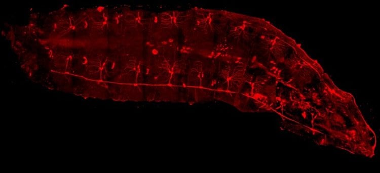
Image Source: NeuroscienceNews.com image is credited to Melissa Rolls, Penn State.
Video Source: The video is credited to Huck Institutes of the Life Sciences
Original Research: Full open access research for “Bilaterian Giant Ankyrins Have a Common Evolutionary Origin and Play a Conserved Role in Patterning the Axon Initial Segment” Timothy Jegla, Michelle M. Nguyen, Chengye Feng, Daniel J. Goetschius, Esteban Luna, Damian B. van Rossum, Bishoy Kamel, Aditya Pisupati, Elliott S. Milner, and Melissa M. Rolls in PLOS Genetics. Published online December 2 2016 doi:10.1371/journal.pgen.1006457
[cbtabs][cbtab title=”MLA”]Penn State “Discovery Rewriting the Evolutionary History of the Nervous System.” NeuroscienceNews. NeuroscienceNews, 8 March 2017.
<https://neurosciencenews.com/evolution-nervous-system-6215/>.[/cbtab][cbtab title=”APA”]Penn State (2017, March 8). Discovery Rewriting the Evolutionary History of the Nervous System. NeuroscienceNew. Retrieved March 8, 2017 from https://neurosciencenews.com/evolution-nervous-system-6215/[/cbtab][cbtab title=”Chicago”]Penn State “Discovery Rewriting the Evolutionary History of the Nervous System.” https://neurosciencenews.com/evolution-nervous-system-6215/ (accessed March 8, 2017).[/cbtab][/cbtabs]
Abstract
Bilaterian Giant Ankyrins Have a Common Evolutionary Origin and Play a Conserved Role in Patterning the Axon Initial Segment
In vertebrate neurons, the axon initial segment (AIS) is specialized for action potential initiation. It is organized by a giant 480 Kd variant of ankyrin G (AnkG) that serves as an anchor for ion channels and is required for a plasma membrane diffusion barrier that excludes somatodendritic proteins from the axon. An unusually long exon required to encode this 480Kd variant is thought to have been inserted only recently during vertebrate evolution, so the giant ankyrin-based AIS scaffold has been viewed as a vertebrate adaptation for fast, precise signaling. We re-examined AIS evolution through phylogenomic analysis of ankyrins and by testing the role of ankyrins in proximal axon organization in a model multipolar Drosophila neuron (ddaE). We find giant isoforms of ankyrin in all major bilaterian phyla, and present evidence in favor of a single common origin for giant ankyrins and the corresponding long exon in a bilaterian ancestor. This finding raises the question of whether giant ankyrin isoforms play a conserved role in AIS organization throughout the Bilateria. We examined this possibility by looking for conserved ankyrin-dependent AIS features in Drosophila ddaE neurons via live imaging. We found that ddaE neurons have an axonal diffusion barrier proximal to the cell body that requires a giant isoform of the neuronal ankyrin Ank2. Furthermore, the potassium channel shal concentrates in the proximal axon in an Ank2-dependent manner. Our results indicate that the giant ankyrin-based cytoskeleton of the AIS may have evolved prior to the radiation of extant bilaterian lineages, much earlier than previously thought.
“Bilaterian Giant Ankyrins Have a Common Evolutionary Origin and Play a Conserved Role in Patterning the Axon Initial Segment” Timothy Jegla, Michelle M. Nguyen, Chengye Feng, Daniel J. Goetschius, Esteban Luna, Damian B. van Rossum, Bishoy Kamel, Aditya Pisupati, Elliott S. Milner, and Melissa M. Rolls in PLOS Genetics. Published online December 2 2016 doi:10.1371/journal.pgen.1006457


