

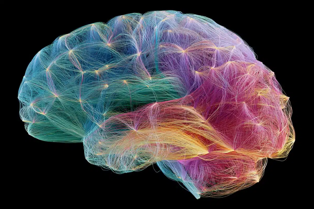
Summary: New research utilized advanced, simultaneous EEG-fMRI tracking to prove that the brain does not run a single process. Instead, the human connectome coordinates multiple entirely separate, asynchronous streams of information processing that run in parallel, opening a new window into clinical neuro-diagnostics and cognitive complexity.
Key Facts
- The Single-Process Myth Debunked: Rather than showing that fMRI signals are merely a sluggish carbon copy of fast electrical brain waves, the concurrent data proved that the brain operates multiple distinct, coordinated streams of information processing at the exact same time.
- The Language Analogy: Dr. Jun compares this asynchronous processing to how the mind decodes speech: the brain simultaneously tracks the rapid flicker of individual phonetic sounds (fast stream), the slower assembly of words (medium stream), and the overarching narrative thread of meaning (slow stream)—all on parallel, independent tracks.
- Asynchronous Blueprints: Remarkably, these separate information streams are built from identical spatial blueprints and tend to unfold across the exact same anatomical brain networks in the same order, yet they operate completely asynchronously.
- The Technical Cross-Artifact Triumph: Recording clean EEG data inside a high-powered, vibrating magnetic MRI machine was a massive hurdle that took years to solve. The team spent nearly half a decade developing specialized safety protocols and data-cleaning algorithms to remove severe magnetic and physical artifacts without destroying vital neurological data.
- Resurrecting EEG’s Clinical Standalone Value: By proving that EEG captures unique, high-fidelity connectome data completely independent of fMRI, this study validates the use of standalone EEG in clinical settings. This is a game-changer for low-income clinics or patients who cannot undergo MRI scans due to metal implants, claustrophobia, or financial barriers.
- Translational Potential for Autoimmune and Neurodegenerative Diseases: Unveiling these asynchronous parallel processing streams provides an entirely new template to study psychiatric and neurological conditions, offering a fresh diagnostic angle to track how dementia, aging, or autoimmune conditions like HIV systematically disrupt the brain’s timing networks.
Source: Beckman Institute
Postdoctoral researcher Suhnyoung Jun has always been interested in brains.
Jun is the co-first author of a recent paper that centers on the connectome, the comprehensive neural network within the brain. She works in the CONNECTlab with psychology professor Sepideh Sadaghiani.
Working with concurrent electroencephalogram and functional magnetic resonance imaging technology at the Beckman Institute’s Biomedical Imaging Center, Jun and her colleagues investigated how the connectome’s dynamics unfold across different timescales, captured by these two technologies at the same time.
In her previous research, Jun focused on heritable features, or traits passed from parents to children through DNA, centering on how patterns of communication within the connectome change over time. For this research, she used either fMRI or EEG data. At the time, she was analyzing each modality separately.
fMRI captures brain activity indirectly through changes in blood oxygenation at slow timescales, while EEG measures electrophysiological signals at much faster timescales. For years, many assumed both modalities were capturing the same underlying brain activity at different speeds, with fMRI simply a slowed-down version of the EEG signal.
Jun and her colleagues challenged this assumption in their recent publication. By recording EEG-fMRI data at the same time, they found the brain isn’t running a single process. Instead, it runs many distinct coordinated processes at the same time. These separate, yet parallel, streams have their own character and unfold independently of each other.
“It’s like when we process language,” she said. “The brain tracks the rapid flicker of individual sounds, the slower arrival of words and the still slower thread of meaning all at once, each on its own stream.”
Access to the Beckman Institute’s facilities was invaluable in conducting this research, Jun said, adding that the research couldn’t have been conducted anywhere else.
“Recording both modalities at the same time was the only way to answer this kind of question,” she said.
Still, the research process was long, spanning almost five years from conception to publication. The team of researchers from the CONNECTlab spent time completing safety training and experimenting, which led to a separate publication on that topic.
In addition, identifying and cleaning artifacts from concurrently recorded EEG and fMRI data took years. Thanks to the staff at the Biomedical Imaging Center, especially BIC Technical Director Brad Sutton, MRI technologist Holly Keleher and a team of grad students in the lab, they eventually found a way to record clean data.
This work began when co-first author Thomas Alderson won a Faculty Early Career Development grant from the National Science Foundation. Sadaghiani contacted previous collaborators to use a set of concurrent EEG-fMRI data from Paris and Jun established a relationship with the Minnesota Center for Twin and Family Research to work with a large EEG dataset of 443 participants. Both these datasets provided additional validation for the findings.
With all these resources and collaborators in place, the team’s results were clear.
“There are multiple streams going on there, but they can talk to each other,” Jun said. “What’s striking is that these separate streams are built from the same spatial blueprints and they tend to play out in the same order, but asynchronously.”
The paper sheds light on the complexity of the brain and its processes and definitively shows that fMRI and EEG capture different information. Jun hopes the impact will go beyond the scientific community. For example, the paper illustrates the usefulness of EEG technology, possibly paving the way for more clinical use in circumstances where patients can’t have MRI scans, either due to expense or the limitations of magnetic MRI technology
“We are losing all their data,” Jun said, “and the MRI-based story that’s going out into the world won’t represent that population.”
This work has the potential to advance research on neurological and psychiatric conditions, including neurodegenerative diseases such as dementia and autoimmune diseases like HIV.
“I hope that my research will help more patients,” Jun said, “and that it will be used by someone who is near to translational work.”
Key Questions Answered:
A: It came down to a lack of technology capable of running both tests cleanly at the same time. Because fMRI scans are slow and look at blood flow, while EEG scans are blindingly fast and look at electricity, scientists logically assumed they were looking at the same movie at different frame rates. They figured blood flow simply lagged behind the electrical activity. By developing a way to safely record both inside the same machine without the giant magnets ruining the electrical signals, the Beckman Institute proved that they are actually recording entirely separate, independent stories playing out over the same neural highways.
A: Think of it like a massive musical orchestra where the brass section, the strings, and the percussion are all playing from the exact same sheet music, but they are all playing at completely different tempos and starting at different times. The physical pathways and the order in which the brain areas fire are identical across both slow and fast speeds, but they operate completely independently of one another. One stream doesn’t wait for or copy the other; they handle different layers of cognitive data concurrently.
A: Currently, the most prominent models of brain health and cognitive aging are built almost exclusively on expensive fMRI data. Dr. Suhnyoung Jun points out that by excluding patients who can’t get MRIs, due to cost, geographic location, or safety issues like pacemakers and metal shrapnel, our medical models are biased and missing huge chunks of the human population. Now that this study proves EEG holds its own unique, independent treasure trove of connectome data, doctors can confidently use standalone, low-cost EEG technology to diagnose and map brain disorders in underserved communities worldwide.
Editorial Notes:
- This article was edited by a Neuroscience News editor.
- Journal paper reviewed in full.
- Additional context added by our staff.
About this connectome research news
Author: Alejandra Pires
Source: Beckman Institute
Contact: Alejandra Pires – Beckman Institute
Image: The image is credited to Neuroscience News
Original Research: Open access.
“Shared spatial and temporal principles govern connectome dynamics across timescales” by Anne-Lise Giraud, Jeremy Harper, Jonathan Wirsich, Maximillian Kirichenko Egan, Parham Mostame, Samar Wagih ElSayed, Sanmi Koyejo, Sepideh Sadaghiani, Sophia A. Giakas, Stephen M. Malone, Suhnyoung Jun, Thomas H. Alderson, William G. Iacono. PNAS
DOI:10.1073/pnas.2535464123
Abstract
Shared spatial and temporal principles govern connectome dynamics across timescales
While the brain processes information at various speeds, little is known about how the functional connectome can concurrently support multiple speeds in parallel. FMRI and electrophysiological modalities have been used to study connectome dynamics at slow and fast speeds, respectively.
But it is often implicitly assumed that these modalities capture the same underlying neural processes, with fMRI doing so through a low-pass temporal filter.
However, recent work suggests the alternative possibility that connectome dynamics comprise distinct processes operating at multiple timescales. If such multiscale connectivity processes indeed coexist, a key question is whether their patterns and sequences are organized on the basis of shared regularities, i.e., common spatial and temporal principles.
In simultaneous human fMRI and source-localized EEG, we investigated the connectome’s foundational constituents—the instantaneous coactivation patterns—across six timescales of neural activity, from infraslow through γ-band. We found streams of recurrent coactivation patterns, or states, that operate in parallel and asynchronously across timescales, thereby forming a timescale-overarching spatial principle. These states also occurred in highly similar sequences at all timescales, revealing a timescale-overarching temporal principle.
Together, these findings indicate that the connectome comprises multiple dynamic streams operating in parallel at distinct speeds, from tens of milliseconds to seconds, rather than a single stream filtered by each modality’s temporal resolution. Spatial and temporal principles that span these streams enable their integration into a unified system.
Consequently, research on human behavior and mental disorders should account for the full range of the connectome’s timescales.


