Knowing how these circuits work could lead to targeted drug therapies for overeating.
Researchers investigating eating disorders often study chemical and neurological functions in the brain to discover clues to overeating. Understanding non-homeostatic eating — or eating that is driven more by palatability, habit and food cues — and how it works in the brain may help neuroscientists determine how to control cravings, maintain healthier weights and promote healthier lifestyles. Scientists at the University of Missouri recently discovered the chemical circuits and mechanisms in the brain that separate food consumption from cravings. Knowing more about these mechanisms could help researchers develop drugs that reduce overeating.
“Non-homeostatic eating can be thought of as eating dessert after you’ve eaten an entire meal,” said Kyle Parker, a former grad student and investigator in the MU Bond Life Sciences Center. “I may know that I’m not hungry, but this dessert is delicious so I’m going to eat it anyway. We’re looking at what neural circuitry is involved in driving that behavior.”
Matthew J. Will, an associate professor of psychological sciences in the MU College of Arts and Science, a research investigator in the Bond Life Sciences Center and Parker’s adviser, says for behavior scientists, eating is described as a two-step process called the appetitive and consummatory phases.
“I think of the neon sign for a donut shop — the logo and the aroma of warm glazed donuts are the environmental cues that kick start the craving, or appetitive, phase,” Will said. “The consummatory phase is after you have that donut in hand and eat it.”
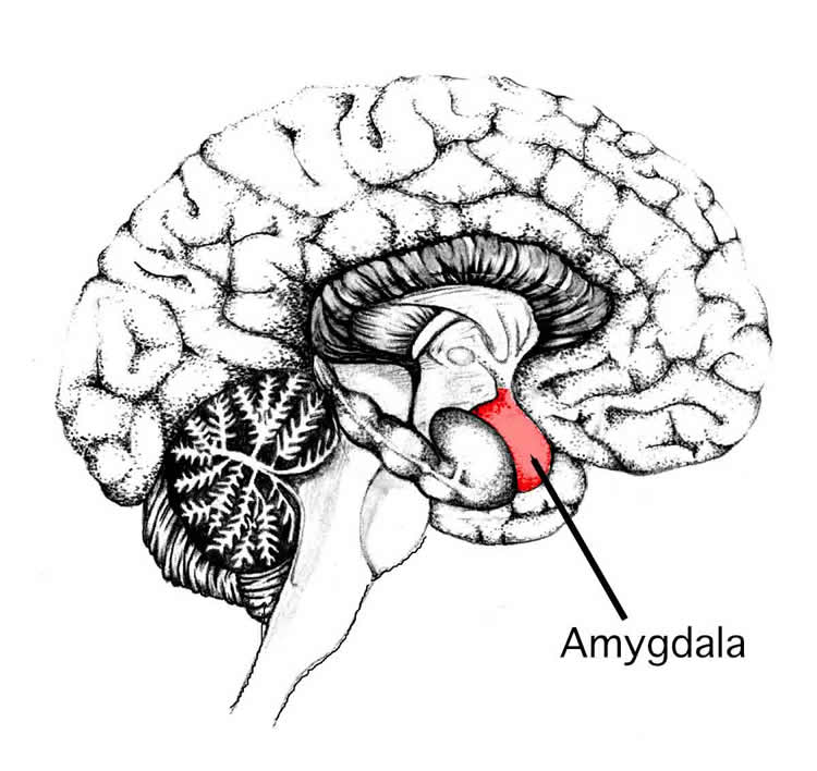
Parker studied the behavior patterns of laboratory rats by activating the brain’s pleasure center, a hotspot in the brain that processes and reinforces messages related to reward and pleasure. He then fed the rats a cookie dough-like diet to exaggerate their feeding behaviors and found that the rats ate twice as much as usual. When he simultaneously inactivated another part of the brain called the basolateral amygdala, the rats stopped binge eating. They kept returning to their food baskets in search of more, but only consumed a normal amount.
“It seemed as if the rats still craved the dough,” Will said. “They kept going back for food but simply didn’t eat. We found that we had interrupted the part of the brain that’s specific to feeding — the circuit attached to actual eating — but not the craving. In essence, we left that craving intact.”
To find out what was happening in the brain during cravings, Parker set up a spin-off experiment. Like before, he switched on the region of the brain associated with reward and pleasure and inactivated the basolateral amygdala in one group of rats but not the other. This time, however, he limited the amount of the high fat diet the rats had access to so that both groups ate the same amount.
Outwardly, both groups of rats displayed the same feeding behaviors. They ate a portion of food, but kept going back and forth to their food baskets. However, inside the brain, Parker saw clear differences. Rats with activated nucleus accumbens showed increased dopamine neuron activity, which is associated with motivated approach behavior.
The team also found that the state of the basolateral amygdala had no effect on dopamine signaling levels. However, in a region of the brain called the hypothalamus, Parker saw elevated levels of orexin-A, a molecule associated with appetite, only in rats with activated basolateral amygdala.
“We showed that what could be blocking the consumption behavior is this block of the orexin behavior,” Parker said.
“The results reinforced the idea that dopamine is involved in the approach — or the craving phase — and orexin-A in the consumption,” Will said.
The team believes that these findings could lead to a better understanding of the different aspects of overeating and drug addiction. By revealing the independent circuitry of craving vs. the actual consumption or drug taking, this could lead to potential drug treatments that are more specific and have less unwanted side effects.
Parker and Will’s study, “Neural activation patterns underlying basolateral amygdala influence on intra-accumbens opioid-driven consummatory versus appetitive high-fat feeding behaviors in the rat,” recently was published in Behavioral Neuroscience. Research was funded in part by the National Institute of Drug Abuse (DA024829). The content is solely the responsibility of the authors and does not necessarily represent the official views of the funding agency.
Funding: Research was funded in part by the National Institute of Drug Abuse (DA024829). The content is solely the responsibility of the authors and does not necessarily represent the official views of the funding agency.
Source: Jeff Sossamon – University of Missouri
Image Credit: The image is in the public domain.
Original Research: Abstract for “Neural activation patterns underlying basolateral amygdala influence on intra-accumbens opioid-driven consummatory versus appetitive high-fat feeding behaviors in the rat” by Parker, Kyle E.; McCabe, Matt P.; Johns, Howard W.; Lund, Dane K.; Odu, Fiona; Sharma, Rishi; Thakkar, Mahesh M.; Cornelison, D. D. W.; and Will, Matthew J. in Behavioral Neuroscience. Published online December 2015 doi:10.1037/bne0000095
Abstract
Neural activation patterns underlying basolateral amygdala influence on intra-accumbens opioid-driven consummatory versus appetitive high-fat feeding behaviors in the rat
The present study explored the role of the amygdala in mediating a unique pattern of feeding behavior driven by intra-accumbens (intra-Acb) opioid activation in the rat. Temporary inactivation of the basolateral amygdala (BLA), via GABAA agonist muscimol administration prevents increased consumption following intra-Acb opioid administration of the selective μ-opioid agonist D-Ala2, NMe-Phe4, Glyol5-enkephalin (DAMGO), yet leaves food approach behaviors intact, particularly after consumption has ended. One interpretation is that inactivation of the BLA selectively blocks neural activity underlying DAMGO-driven consummatory (consumption) but not appetitive (approach) behaviors. The present experiments take advantage of this temporal dissociation of consumption and approach behaviors to investigate their associated neural activity. Following either intra-Acb saline or DAMGO administration, with or without BLA muscimol administration, rats were given 2-hr access to a limited amount of high-fat diet. Immediately following the feeding session, rats were sacrificed and brains assayed for neural activity patterns across critical brain regions known to regulate both appetitive and consummatory feeding behaviors. The results show that intra-Acb DAMGO administration increased c-Fos activation in orexin neurons within the perifornical area of the hypothalamus and that this increase in activation is blocked by BLA muscimol inactivation. Intra-Acb DAMGO administration significantly increased c-Fos activation within dopaminergic neurons of the ventral tegmental area, compared to saline controls, and BLA inactivation had no effect on this increase. Overall, these data provide underlying circuitry that may mediate the selective influence of the BLA on driving consummatory, but not appetitive, feeding behaviors in a model of hedonically driven feeding behavior.
“Neural activation patterns underlying basolateral amygdala influence on intra-accumbens opioid-driven consummatory versus appetitive high-fat feeding behaviors in the rat” by Parker, Kyle E.; McCabe, Matt P.; Johns, Howard W.; Lund, Dane K.; Odu, Fiona; Sharma, Rishi; Thakkar, Mahesh M.; Cornelison, D. D. W.; and Will, Matthew J. in Behavioral Neuroscience. Published online December 2015 doi:10.1037/bne0000095