

Summary: Based on the actions of Arc, when one synapse strengthens, the neighboring synapses weaken, a new study reveals.
Source: MIT.
Our brains are famously flexible, or “plastic,” because neurons can do new things by forging new or stronger connections with other neurons. But if some connections strengthen, neuroscientists have reasoned, neurons must compensate lest they become overwhelmed with input. In a new study in Science, researchers at the Picower Institute for Learning and Memory at MIT demonstrate for the first time how this balance is struck: when one connection, called a synapse, strengthens, immediately neighboring synapses weaken based on the action of a crucial protein called Arc.
Senior author Mriganka Sur said he was excited but not surprised that his team discovered a simple, fundamental rule at the core of such a complex system as the brain, where 100 billion neurons each have thousands of ever-changing synapses. He likens it to how a massive school of fish can suddenly change direction, en masse, so long as the lead fish turns and every other fish obeys the simple rule of following the fish right in front of it.
“Collective behaviors of complex systems always have simple rules,” said Sur, Paul E. and Lilah Newton Professor of Neuroscience in the Picower Institute and the department of Brain and Cognitive Sciences at MIT. “When one synapse goes up, within 50 micrometers there is a decrease in the strength of other synapses using a well-defined molecular mechanism.”
This finding, he said, provides an explanation of how synaptic strengthening and weakening combine in neurons to produce plasticity.
Multiple manipulations
Though the rule they found was simple, the experiments that revealed it were not. As they worked to activate plasticity in the visual cortex of mice and then track how synapses changed to make that happen, lead authors Sami El-Boustani and Jacque Pak Kan Ip, postdoctoral researchers in Sur’s lab, accomplished several firsts.
In one key experiment, they invoked plasticity by changing a neuron’s “receptive field,” or the patch of the visual field it responds to. Neurons receive input through synapses on little spines of their branch-like dendrites. To change a neuron’s receptive field, the scientists pinpointed the exact spine on the relevant dendrite of the neuron, and then closely monitored changes in its synapses as they showed the mouse a target in a particular place on a screen that differed from the neuron’s original receptive field. Whenever the target was in the new receptive field position they wanted to induce, they reinforced the neuron’s response by flashing a blue light inside the mouse’s visual cortex, instigating extra activity just like another neuron might. The neuron had been genetically engineered to be activated by light flashes, a technique called “optogenetics.”
The researchers did this over and over. Because the light stimulation correlated with each appearance of the target in the new position in the mouse’s vision, this caused the neuron to strengthen a particular synapse on the spine, encoding the new receptive field.
“I think it’s quite amazing that we are able to reprogram single neurons in the intact brain and witness in the living tissue the diversity of molecular mechanisms that allows these cells to integrate new functions through synaptic plasticity,” El-Boustani said.
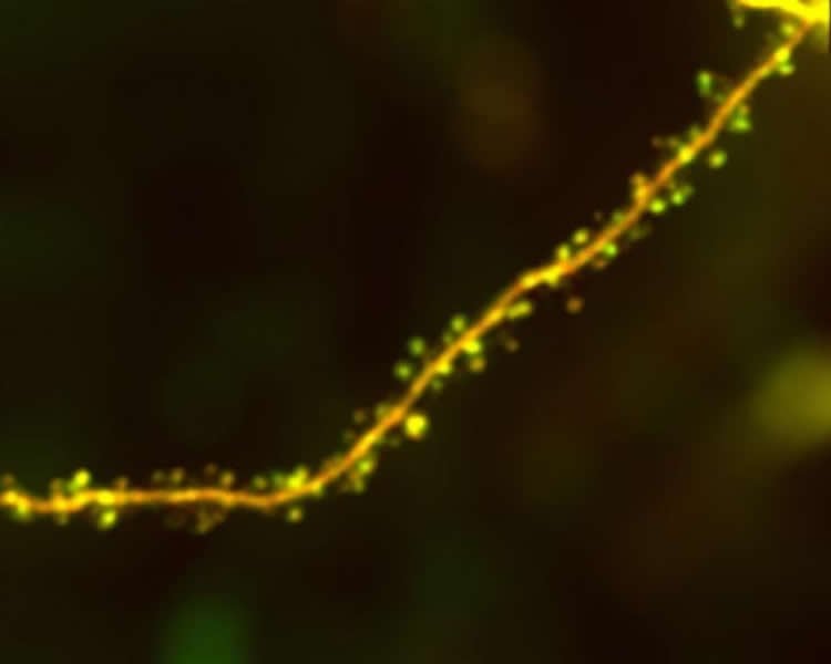
As the synapse for the new receptive field grew, the researchers could see under the two-photon microscope that nearby synapses also shrank. They did not observe these changes in experimental control neurons that lacked the optogenetic stimulation.
But then they went further to confirm their findings. Because synapses are so tiny, they are near the limit of the resolution of light microscopy. So after the experiments the team dissected the brain tissues containing the dendrites of manipulated and control neurons and shipped them to co-authors at the Ecole Polytechnique Federal de Lausanne in Switzerland. They performed a specialized, higher-resolution, 3D electron microscope imaging, confirming that the structural differences seen under the two-photon microscope were valid.
“This is the longest length of dendrite ever reconstructed after being imaged in vivo,” said Sur, who also directs the Simons Center for the Social Brain at MIT.
Of course, reprogramming a mouse’s genetically engineered neuron with flashes of light is an unnatural manipulation, so the team did another more classic “monocular deprivation” experiment in which they temporarily closed one eye of a mouse. When that happens synapses in neurons related to the closed eye weaken and synapses related to the still open eye strengthen. Then when they reopened the previously closed eye, the synapses rearrange again. They tracked that action, too, and saw that as synapses strengthen, their immediate neighbors would weaken to compensate.
Solving the mystery of the Arc
Having seen the new rule in effect, the researchers were still eager to understand how neurons obey it. They used a chemical tag to watch how key “AMPA” receptors changed in the synapses and saw that synaptic enlargement and strengthening correlated with more AMPA receptor expression while shrinking and weakening correlated with less AMPA receptor expression.
The protein Arc regulates AMPA receptor expression, so the team realized they had to track Arc to fully understand what was going on. The problem, Sur said, is that no one had ever done that before in the brain of a live, behaving animal. So the team reached out to co-authors at the Kyoto University Graduate School of Medicine and the University of Tokyo, who invented a chemical tag that could do so.
Using the tag, the team could see that the strengthening synapses were surrounded with weakened synapses that had enriched Arc expression. Synapses with reduced amount of Arc were able to express more AMPA receptors whereas increased Arc in neighboring spines caused those synapses to express less AMPA receptors.
“We think Arc maintains a balance of synaptic resources,” Ip said. “If something goes up, something must go down. That’s the major role of Arc.”
Sur said the study therefore solves a mystery of Arc: No one before had understood why Arc seemed to be upregulated in dendrites undergoing synaptic plasticity, even though it acts to weaken synapses, but now the answer was clear. Strengthening synapses increase Arc to weaken their neighbors.
Sur added that the rule helps explain how learning and memory might work at the individual neuron level because it shows how a neuron adjusts to the repeated simulation of another.
Funding: Funding for the research came from the Picower Institute Innovation Fund, The Simons Center for the Social Brain, a Marie Curie postdoctoral fellowship, a Human Frontier Science Program Long-Term Fellowship, The National Institutes of Health (grants: NS090473, EY007023), The National Science Foundation (EF1451125) and KAKENHI (15H04258,15H02358, 17H06312).
Source: David Orenstein – MIT
Publisher: Organized by NeuroscienceNews.com.
Image Source: NeuroscienceNews.com image is credited to Sur, et. al..
Original Research: Abstract for “Locally coordinated synaptic plasticity of visual cortex neurons in vivo” by Sami El-Boustani, Jacque P. K. Ip, Vincent Breton-Provencher, Graham W. Knott, Hiroyuki Okuno, Haruhiko Bito, and Mriganka Sur in Science. Published June 22 2018.
doi:10.1126/science.aao0862
[cbtabs][cbtab title=”MLA”]MIT “Fundamental Rule of Brain Plasticity Discovered.” NeuroscienceNews. NeuroscienceNews, 21 June 2018.
<https://neurosciencenews.com/brain-plasticity-neuroscience-9414/>.[/cbtab][cbtab title=”APA”]MIT (2018, June 21). Fundamental Rule of Brain Plasticity Discovered. NeuroscienceNews. Retrieved June 21, 2018 from https://neurosciencenews.com/brain-plasticity-neuroscience-9414/[/cbtab][cbtab title=”Chicago”]MIT “Fundamental Rule of Brain Plasticity Discovered.” https://neurosciencenews.com/brain-plasticity-neuroscience-9414/ (accessed June 21, 2018).[/cbtab][/cbtabs]
Abstract
Locally coordinated synaptic plasticity of visual cortex neurons in vivo
Plasticity of cortical responses in vivo involves activity-dependent changes at synapses, but the manner in which different forms of synaptic plasticity act together to create functional changes in neurons remains unknown. We found that spike timing–induced receptive field plasticity of visual cortex neurons in mice is anchored by increases in the synaptic strength of identified spines. This is accompanied by a decrease in the strength of adjacent spines on a slower time scale. The locally coordinated potentiation and depression of spines involves prominent AMPA receptor redistribution via targeted expression of the immediate early gene product Arc. Hebbian strengthening of activated synapses and heterosynaptic weakening of adjacent synapses thus cooperatively orchestrate cell-wide plasticity of functional neuronal responses.


