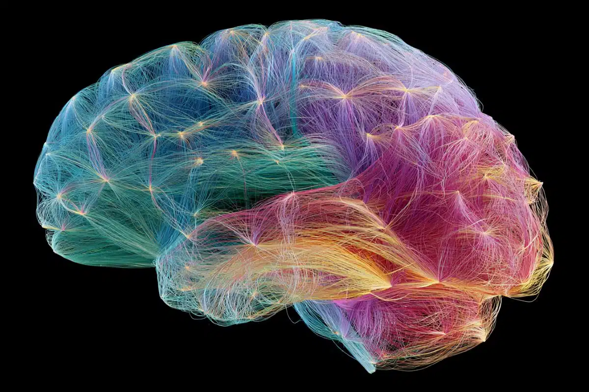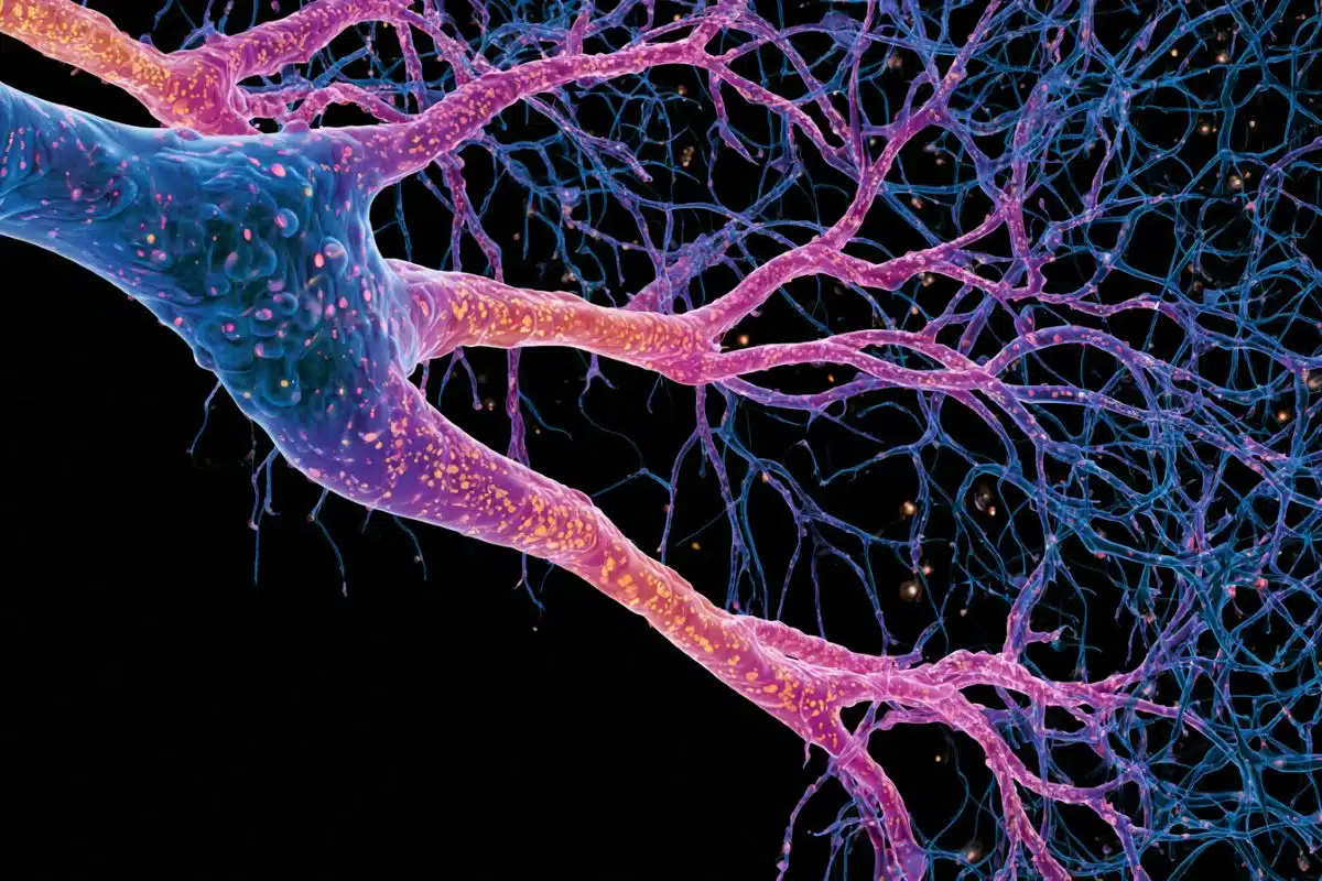
Summary: Researchers discovered that axon generation is actually an autonomous, internally driven process. The team proved that young nerve cells utilize an internal protein complex to methodically “unzip” their own structural scaffolding from the inside out, establishing early brain wiring via an intrinsic genetic protocol.
Key Facts
- The Intrinsic Control Shift: Rather than being passively shaped by external biological chemical trails, embryonic neurons actively remodel their own cytoskeleton (the cell’s internal structural scaffold) to trigger axon outgrowth, originating from the cell body (soma).
- The Dance of the Neurites: Early symmetric neurons exhibit small, bud-like extensions called neurites. These extensions display a highly rhythmic “two steps forward, one step back” movement behavior, constantly stretching outward and shrinking backward on a minute-by-minute basis.
- The Arp2/3 Molecular Zipper: The key to breaking this loop is a specialized protein complex called Arp2/3. Acting like a microscopic molecular zipper, Arp2/3 locally opens the cell’s tight, tension-bearing structural “corset,” allowing individual neurites to bulge outward.
- Wave-Like Propagation: This internal unzipping action travels outward through a single neurite at a time like a physical wave. The wave continues until it meets the mechanical resistance of the remaining cellular corset, which forces the neurite back into a brief rest state.
- Microtubule Lock-In: While these random, wave-driven expansions alternate between different neurites by chance, a parallel internal process is underway: rigid structural proteins (microtubules) grow outward from within. Eventually, one lucky neurite accumulates enough rigid microtubule scaffolding to resist pulling back.
- Symmetry Shattered: Once this stable tipping point is reached within roughly 48 hours, that specific neurite transitions into independent, rapid growth to become the official axon. The overall wave-driven shape-shifting stops, and the remaining neurites are locked into becoming input-receiving dendrites.
Source: DZNE
Neurons in the brain and spinal cord form a vast network in which each cell receives many inputs but sends output through only a single, long extension: the “axon”.
“If our neurons had multiple axons, this would cause chaos in the brain,” says Professor Frank Bradke, a neurobiologist and research group leader at DZNE.
“Nature has therefore found a clever way to make sure that neurons generate only one axon. This applies not only to humans, but across the entire animal kingdom. So, we’re dealing with very fundamental processes that shape the wiring of the brain and nervous system.”
Breaking symmetry
During early embryonic development, neurons are initially largely symmetric, exhibiting small projections known as neurites. E
ventually one of these develops into the axon, thereby breaking the symmetry. Until now, it was largely assumed that this process is determined by biological growth factors that act on a neuron from the outside – and that, much like attractants, lead to the development of the axon. The team led by Frank Bradke reaches a different conclusion.
“According to our observations, the axon forms as a result of a remodeling of the cytoskeleton initiated by the young neuron. The process originates in the cell body, the so-called soma – the very center of the neuron,” says Dr. Tien-chen Lin, first author of the current publication and a scientist at DZNE.
Young neurons display a rhythmic behavior: Their neurites stretch out somewhat, and then shrink back slightly.
“This happens on a minute-by-minute basis. In a sense, the process follows the principle of two steps forward and one step back. This sequence repeats again and again,” says Tien-chen Lin.
Within typically 48 hours, however, one of the neurites grows into an axon. The remaining neurites later develop into receptors for inputs.
“Actually, this recurring process was already known. But it was unclear what lies behind it. We have now been able to shed considerable light on the underlying mechanisms.”
The key lies in the neuron’s cytoskeleton, a tension‑bearing, molecular scaffold that acts like a corset around the cell.
“This is where a protein complex called Arp2/3 enters the picture. Our findings show that it works like a zipper, locally opening the cell’s corset,” says Tien-chen Lin. “By doing so, Arp2/3 drives the cell’s rhythmic shape-shifting, repeatedly loosening a network that would otherwise tighten up again.”
Wave-like propagation
The researchers found that Arp2/3 always acts on only one neurite at a time, temporarily enabling its growth.
“This comes with a locally confined restructuring of the cytoskeleton that spreads like a wave,” says Tien-chen Lin. “These events continue until the wave subsides because, although the cellular corset has been loosened, it still offers a certain degree of resistance. Then the process begins again. The same neurite may be affected once more, or a different one may be involved. Which one seems to be a matter of chance.”
Parallel to this outward extension, relatively rigid structural proteins grow into the neurites from within.
“Eventually, one of the neurites becomes stable enough to resist being pulled back. It can then continue growing independently of Arp2/3 and ultimately develops into the axon. Meanwhile, the overall ’wave-driven’ outgrowth comes to a stop,” says Tien-chen Lin.
Open questions
“We cannot exclude the possibility that external factors play a certain role. However, given our data, we are convinced that the basic process that drives axon growth originates within the cell itself,” says Frank Bradke.
Open questions remain: What initiates the remodeling? Why does it proceed rhythmically and one neurite at a time? And, why does remodeling stop as soon as one of the neurites has grown large enough?
“The young nerve cell presumably follows a protocol encoded in its genome. However, we do not yet know the relevant genetic program, and our understanding of the associated regulatory processes remains limited. Thus, there is still plenty of research ahead, which motivates us to continue pursuing this topic.”
Key Questions Answered:
A: Think of a neuron like an ultra-precise telephone line. It is biologically designed to collect information from thousands of neighbors through its many input roots (dendrites), but it must broadcast its finalized message through a single, dedicated output wire (the axon). If a neuron sprouted multiple axons, it would blast its electrical signals into multiple unintended circuits simultaneously, causing widespread cross-talk, short-circuiting sensory loops, and destroying the brain’s delicate computational symmetry.
A: Every young neuron is wrapped in a highly tense, rigid mesh network of structural proteins that acts like a cellular corset, keeping the cell tight and round. Dr. Tien-chen Lin discovered that the Arp2/3 complex acts as a localized release valve. When it activates at the base of a neurite, it unzips the interlocking threads of that corset, temporarily loosening the tension. This allows the internal contents of the cell to push outward in a wave, extending that specific branch further into space.
A: According to the DZNE data, the initial selection process is actually driven by chance. The Arp2/3 complex randomly shifts from one neurite to another, causing them all to rhythmically stretch and contract. However, as these branches take turns expanding, rigid rod-like structural proteins called microtubules are continuously growing outward from the center. Eventually, purely by coincidence, one neurite remains expanded just long enough for these rigid rods to pack inside and lock its skeleton in place, permanently stabilizing it so it can no longer shrink back.
Editorial Notes:
- This article was edited by a Neuroscience News editor.
- Journal paper reviewed in full.
- Additional context added by our staff.
About this neuroscience research news
Author: Marcus Neitzert
Source: DZNE
Contact: Marcus Neitzert – DZNE
Image: The image is credited to Neuroscience News
Original Research: Open access.
“An intrinsic cytoskeletal oscillator establishes neuronal polarity” by Tien-chen Lin (林天正), Charlotte H. Coles, Eissa Alfadil, Florian Fäßler, Andreas Husch, Sebastian Dupraz, Thorben Pietralla, Akihiro Narita, Max Schelski, Kevin C. Flynn, Sina Stern, Christoph Möhl, Brett J. Hilton, Franz Vauti, Hans-Henning Arnold, Florian K. M. Schur & Frank Bradke. Nature
DOI:10.1038/s41586-026-10755-6
Abstract
An intrinsic cytoskeletal oscillator establishes neuronal polarity
Neurons acquire polarity by specifying one neurite as the axon, whereas the others become dendrites. But how this fundamental asymmetry is established remains unclear. Neuronal polarization has been thought to rely primarily on growth cones that sense external cues.
Here we show that growth cones alone do not direct this process and that the soma acts as a central organizer of neuronal polarization. Using live imaging and genetic loss-of-function approaches in vivo, combined with optogenetic control and local cytoskeletal perturbations in cultured neurons, we uncover a soma-initiated oscillatory program that primes axon selection.
Periodic actin branching that depends on the actin-related protein 2/3 (ARP2/3) complex at the soma remodels a global actomyosin network, thereby generating an actin wave that retracts neurites before propagating into a single neurite tip. Exposure to this wave relaxes local actomyosin contractility, which drives a transient microtubule-based protrusion and biases this neurite towards axon fate.
As the cell exits this oscillatory stage, this neurite can overcome global inhibition and extend independently of ARP2/3, whereas actomyosin activity suppresses axon formation in the remaining neurites so that they subsequently become dendrites. This soma-driven mechanism ensures the emergence of a single axon independent of environmental cues and underpins the unidirectional information flow in neuronal circuits.