

Summary: Researchers use a newly developed imaging technique that makes tissue transparent to visualize brain tissue from deceased patients with Alzheimer’s disease.
Source: Cell Press.
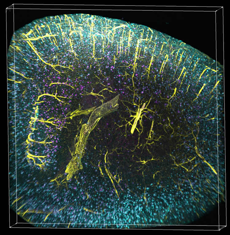
Rockefeller University researchers have used a recently-developed imaging technique that makes tissue transparent to visualize brain tissue from deceased patients with Alzheimer’s disease, exposing nonrandom, higher-order structures of beta amyloid plaques–sticky clumps of a toxic protein typically found in the brains of people with Alzheimer’s. The findings appear July 14 in Cell Reports.
“Until now, we’ve been studying the brain using 2D slices; and I’ve always felt that was inadequate, because it’s a complex, 3D structure with many interlocking components,” says senior author Marc Flajolet, an assistant professor in the Laboratory of Molecular and Cellular Neuroscience at The Rockefeller University. “Not only was slicing time consuming and 3D reconstruction laborious when not erroneous, it gave us a limited view. We needed some way to look at this 3D structure in all of its dimensions without preliminary slicing of the brain.”
The researchers wanted to go beyond the traditional 3D brain imaging (e.g., PET or fMRI scans), which show brain activity in a broad way, but have a low resolution overall. To circumvent this, the research team turned to a recently-developed method, called “iDISCO.” Here, brains are soaked in a solution that imbue the fats within it with a charge, before being exposed to an electrical field with an opposite charge, which behaves like a magnet, forcing all of the fat out of the brain tissue.
The result, says Flajolet, is a brain that is hard and transparent, almost “like glass,” which allowed the researchers to see the amyloid plaques in full detail and in 3D, in a full mouse brain hemisphere, as well as in small blocks of human brain tissue.
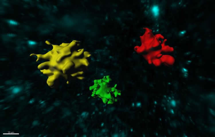
“In mouse models, plaques are rather small, homogenous in size and shape, and not grouped in any specific way,” says Flajolet. “But in the human brain, we were seeing more heterogeneity, larger plaques and these new, complex patterns.” These structures, called TAPs (three dimensional amyloid patterns) may have implications for the future of Alzheimer’s disease treatment, he says. By comparing doctor’s reports of a patient’s symptoms with images of the patient’s brain post-mortem, they may be able to classify different categories of Alzheimer’s disease.
“There are people with brains full of plaques and no dementia at all”, he says, “and there are those with brains free of plaques with many of the symptoms.” In light of that, the way that current clinical trials view the disease–namely, that there is one category–might be incorrect, he says. It’s possible that current drugs may be beneficial only for a subset of Alzheimer patients, but we have no way to distinguish them at this day.
Flajolet stressed that, moving forward, we need a better understanding of these plaques, and Alzheimer’s hallmarks in general, as the relationship between their presence and the severity of the disease is not clear-cut. “Perhaps this will lead to the development of new and better targeted drugs, or allow us to rethink the drugs we have now–that’s what we hope for.”
Funding: This work was supported, in part, by grants from the U.S. National Institutes of Health, the Fisher Center for Alzheimer’s Research Foundation, and the Cure Alzheimer’s Fund. Additional support was received from the Empire State Stem Cell Fund through the New York State Department of Health.
Source: Shoshana Wodinsky – Cell Press
Image Source: This NeuroscienceNews.com images are credited to Dr. Thomas Liebmann, The Rockefeller University.
Original Research: Full open access research for “Three-Dimensional Study of Alzheimer’s Disease Hallmarks Using the iDISCO Clearing Method” by Thomas Liebmann, Nicolas Renier, Karima Bettayeb, Paul Greengard, Marc Tessier-Lavigne, and Marc Flajolet in Cell Reports. Published online July 14 2016 doi:10.1016/j.celrep.2016.06.060
[cbtabs][cbtab title=”MLA”]Cell Press. “Unexpected Arrangement of Plaques in Alzheimer’s Afflicted Brains.” NeuroscienceNews. NeuroscienceNews, 14 July 2016.
<https://neurosciencenews.com/alzheimers-plaque-arrangement-4680/>.[/cbtab][cbtab title=”APA”]Cell Press. (2016, July 14). Unexpected Arrangement of Plaques in Alzheimer’s Afflicted Brains. NeuroscienceNews. Retrieved July 14, 2016 from https://neurosciencenews.com/alzheimers-plaque-arrangement-4680/[/cbtab][cbtab title=”Chicago”]Cell Press. “Unexpected Arrangement of Plaques in Alzheimer’s Afflicted Brains.” https://neurosciencenews.com/alzheimers-plaque-arrangement-4680/ (accessed July 14, 2016).[/cbtab][/cbtabs]
Abstract
Three-Dimensional Study of Alzheimer’s Disease Hallmarks Using the iDISCO Clearing Method
Highlights
•iDISCO clearing is used to detect amyloid plaques in a full mouse brain hemisphere
•3D amyloid patterns (TAPs) are detected in human brain archival samples
•Triple labeling of cleared tissues allows highly contextual analysis of amyloidogenesis
•Automated anatomical mapping empowers accurate and fast quantitation of plaques
Summary
Amyloidosis is a major problem in over one hundred diseases, including Alzheimer’s disease (AD). Using the iDISCO visualization method involving targeted molecular labeling, tissue clearing, and light-sheet microscopy, we studied plaque formation in the intact AD mouse brain at up to 27 months of age. We visualized amyloid plaques in 3D together with tau, microglia, and vasculature. Volume imaging coupled to automated detection and mapping enables precise and fast quantification of plaques within the entire intact mouse brain. The present methodology is also applicable to analysis of frozen human brain samples without specialized preservation. Remarkably, amyloid plaques in human brain tissues showed greater 3D complexity and surprisingly large three-dimensional amyloid patterns, or TAPs. The ability to visualize amyloid in 3D, especially in the context of their micro-environment, and the discovery of large TAPs may have important scientific and medical implications.
“Three-Dimensional Study of Alzheimer’s Disease Hallmarks Using the iDISCO Clearing Method” by Thomas Liebmann, Nicolas Renier, Karima Bettayeb, Paul Greengard, Marc Tessier-Lavigne, and Marc Flajolet in Cell Reports. Published online July 14 2016 doi:10.1016/j.celrep.2016.06.060


