

Summary: A new study could help explain why children born to mothers infected by Zika are more likely to have microcephaly.
Source: Cell Press.
Cranial neural crest cells–which give rise to the bones and cartilage of the skull–are vulnerable to Zika virus, report Stanford University School of Medicine researchers September 29 in Cell Host & Microbe. The discovery, made by infecting in vitro cultures of human cells, offers a potential mechanism for how children born with the virus can have smaller-than-average skulls and disproportionate facial features.
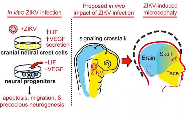
The researchers also found that Zika has slightly different effects on cranial neural crest cells compared to neural progenitor cells, which have received much attention due to their connection with microcephaly. While the virus rapidly kills neural progenitor cells, infection of cranial neural crest cells does not lead to high rates of cell death. Instead, Zika causes them to secrete signaling molecules that promote the formation of new neural cells. In cell culture, elevated levels of these molecules were enough to induce premature differentiation, migration, and death of human neural progenitors.
“In addition to direct effects of Zika virus on neural progenitors and their derivatives, this virus could affect brain development indirectly, through a signaling cross-talk between embryonic cell types,” says co-senior author Joanna Wysocka, a chemical and systems biologist at the Stanford University School of Medicine. “Neural crest cells are one example, but such mechanisms may also be relevant to other tissues that come in contact with the developing brain during head formation and could be infected by Zika virus.”

Wysocka and co-senior author Catherine Blish, a Stanford clinician-scientist, were interested in studying cranial neural crest cells because during embryogenesis they form the majority of bone and cartilage of the head and communicate with the developing brain. They hypothesized that infection of cranial neural crest cells by Zika could cause the disruption in this communication.
“Our in vitro studies raise an intriguing possibility that the Zika virus can infect human cranial neural crest cells in the developing embryo, which in turn could influence brain development through altered paracrine signaling–and also potentially directly affect development of craniofacial structures,” Wysocka says. “As neural crest cell formation occurs during a specific window of embryogenesis (namely, the first trimester, which intriguingly has been correlated with poor birth outcomes in Zika-infected mothers), we do not anticipate similar effects in adults.”
While an interesting line of future study, the authors emphasize that they have no direct proof that Zika virus infects cranial neural crest cells in animals or humans, nor evidence that such infection would be sufficient to result in microcephaly.
Funding: This work was supported by the Stanford Child Health Research Institute and Stanford University School of Medicine, the National Institutes of Health, the March of Dimes Birth Defects Foundation, the Howard Hughes Medical Institute, the National Institute of Dental and Craniofacial Research, the Eunice Kennedy Shriver National Institute of Child Health and Human Development, and a Stanford Systems Biology Seed Grant.
Source: Joseph Caputo – Cell Press
Image Source: This NeuroscienceNews.com images are credited to Rachel Greenberg and Bayless and Greenberg et al./Cell Host & Mircrobe 2016.
Original Research: Full open access research for “Zika Virus Infection Induces Cranial Neural Crest Cells to Produce Cytokines at Levels Detrimental for Neurogenesis” by Nicholas L. Bayless, Rachel S. Greenberg, Tomek Swigut, Joanna Wysocka, and Catherine A. Blish in Cell Host and Microbe. Published online September 29 2016 doi:10.1016/j.chom.2016.09.006
[cbtabs][cbtab title=”MLA”]Cell Press. “Zika Infects and Affects Neural Cells Related to Skull Formation.” NeuroscienceNews. NeuroscienceNews, 3 October 2016.
<https://neurosciencenews.com/skull-formation-zika-5180/>.[/cbtab][cbtab title=”APA”]Cell Press. (2016, October 3). Zika Infects and Affects Neural Cells Related to Skull Formation. NeuroscienceNews. Retrieved October 3, 2016 from https://neurosciencenews.com/skull-formation-zika-5180/[/cbtab][cbtab title=”Chicago”]Cell Press. “Zika Infects and Affects Neural Cells Related to Skull Formation.” https://neurosciencenews.com/skull-formation-zika-5180/ (accessed October 3, 2016).[/cbtab][/cbtabs]
Abstract
Zika Virus Infection Induces Cranial Neural Crest Cells to Produce Cytokines at Levels Detrimental for Neurogenesis
Zika virus (ZIKV) infection during pregnancy is linked to microcephaly, which is attributed to infection of developing brain structures. ZIKV infects neural progenitor cells in vitro, though its effects on other developmentally relevant stem cell populations, including cranial neural crest cells (CNCCs), have not been assessed. CNCCs give rise to most cranial bones and exert paracrine effects on the developing brain. Here, we report that CNCCs are productively infected by ZIKV, but not by the related dengue virus. ZIKV-infected CNCCs undergo limited apoptosis but secrete cytokines that promote death and drive aberrant differentiation of neural progenitor cultures. Addition of two such cytokines, LIF or VEGF, at levels comparable to those secreted by ZIKV-infected CNCCs is sufficient to recapitulate premature neuronal differentiation and apoptotic death of neural progenitors. Thus, our results suggest that CNCC infection by ZIKV may contribute to associated embryopathies through signaling crosstalk between developing face and brain structures.
“Zika Virus Infection Induces Cranial Neural Crest Cells to Produce Cytokines at Levels Detrimental for Neurogenesis” by Nicholas L. Bayless, Rachel S. Greenberg, Tomek Swigut, Joanna Wysocka, and Catherine A. Blish in Cell Host and Microbe. Published online September 29 2016 doi:10.1016/j.chom.2016.09.006


