

Summary:
Researchers have created an unprecedented high-resolution map of the mouse motor cortex, identifying 16 distinct subregions organized across two anatomical axes rather than the traditional two-zone model. The study reveals that the primary and secondary motor cortices operate in parallel rather than hierarchically, offering a standardized anatomical framework to study selective neuronal vulnerability in diseases like ALS and frontotemporal dementia.
Key Facts:
- 16 Distinct Subregions Across Two Axes: Analyzing 547 axonal tracing experiments, researchers categorized the motor cortex into 16 modules arranged in three rows: an anterior-posterior axis separating cognitive planning from sensory integration, and a medial-lateral axis organizing body representations from trunk to mouth.
- Parallel Rather Than Hierarchical Architecture: The primary and secondary motor cortices project in tandem to downstream brainstem and spinal cord targets, challenging classical theories that assumed a top-down structural hierarchy.
- Multimodal Validation & Open Access: The 16-subregion blueprint was independently verified across single-neuron tracing, projection analyses, and cell-type profiling, and has been integrated into the open-source BrainGlobe atlas for global research.
Source: University of Basel / Allen Institute / Friedrich Miescher Institute for Biomedical Research
From writing words and playing catch to chewing food, the mammalian motor cortex is the central hub directing voluntary movement. Yet for decades, neuroscience relied on a coarse understanding of its internal architecture. Most standard reference atlases divided the area broadly into two generalized compartments: the primary motor cortex (M1) and the secondary motor cortex (M2).
However, this two-part division failed to explain the sophisticated functional specialization, diverse cellular populations, and intricate connection topographies that drive motor execution.
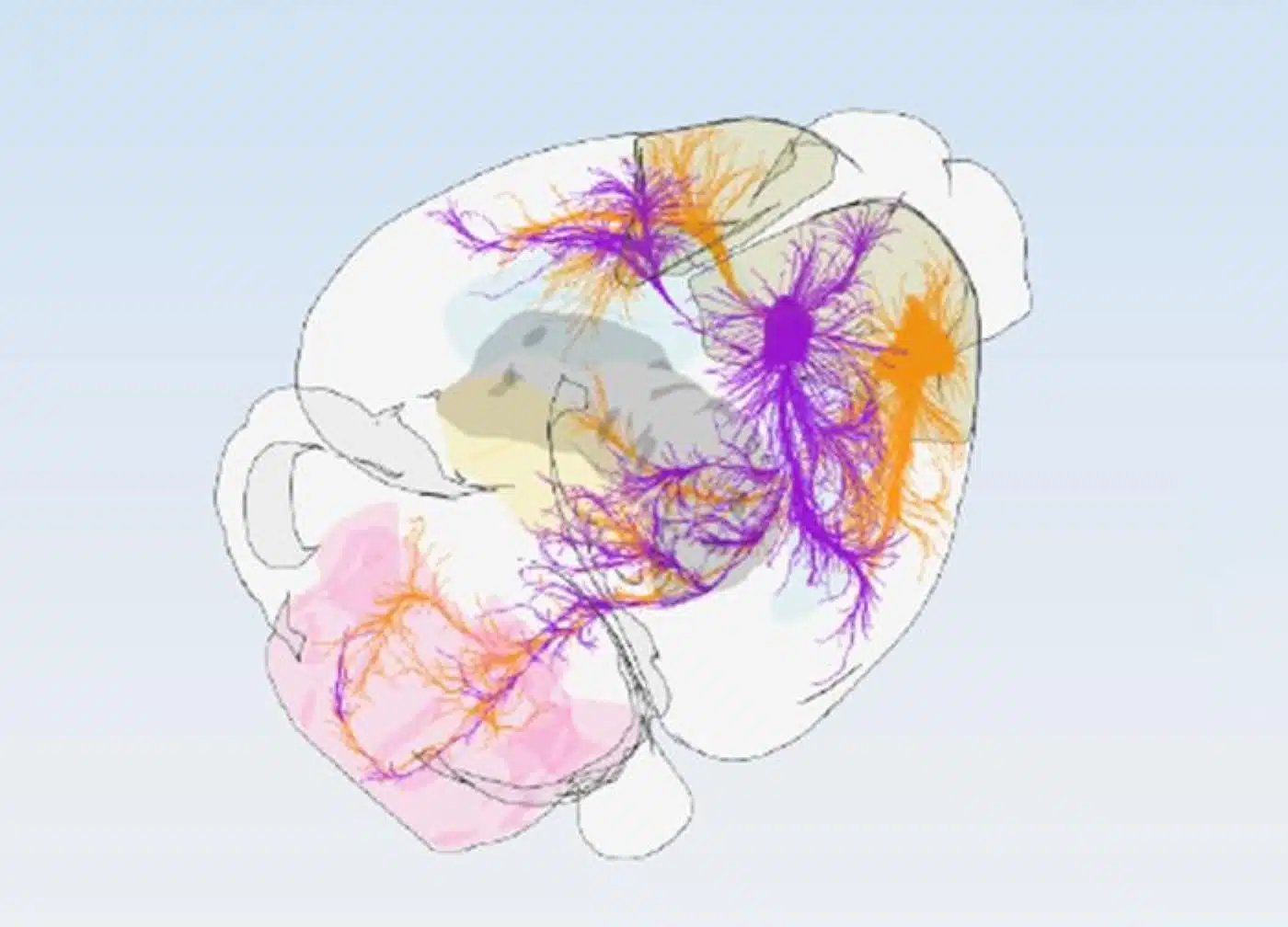
Now, an international collaboration between the University of Basel, the Friedrich Miescher Institute for Biomedical Research (FMI), and the Allen Institute has overhauled this traditional model. By examining how outgoing signals traverse the brain, the team mapped the mouse motor cortex into 16 functionally distinct subregions, each defined by an exclusive wiring pattern.
“Understanding the precise wiring of the brain is essential for developing effective treatments for brain diseases. Here, our Swiss collaborators’ expert charting of the functionally specific motor cortex circuit, combined with the Allen Institute’s foundational connectivity atlas, resulted in such a precision map that drives movement control,” said Hongkui Zeng, Executive Vice President and Director of Brain Science at the Allen Institute and senior author of the study.
A Dual-Axis Coordinate System: 16 Motor Modules
To assemble the map, researchers evaluated 547 individual projection-tracing datasets from the Allen Institute’s Mouse Brain Atlas, systematically cataloging where microscopic cortical coordinates send signals across sensory, motor, and cognitive brain targets.
By clustering areas that share target destinations, the team discovered that the motor cortex is arranged into 16 discrete modules organized into three rows along two primary biological axes:
- Anterior-to-Posterior Axis: Divides regions involved in high-level motor planning and decision-making (anterior) from those coupled directly with sensory feedback, such as proprioception and touch (posterior).
- Medial-to-Lateral Axis: Organizes somatotopic body maps, transitioning from the trunk and limbs on one side to the jaw, mouth, and face on the other.
To confirm the 16-region architecture was biologically consistent, the researchers validated it using two independent methodologies: reconstructing the morphology of individual projecting neurons and profiling the spatial distribution of diverse cortical cell types. All three approaches converged on the identical 16-subregion layout.
Additionally, the investigators resolved a long-standing debate concerning cortical hierarchy. They discovered that primary and secondary motor regions project in tandem directly into the brainstem and spinal cord, refuting the assumption that M2 strictly relays commands through M1 in a rigid hierarchy.
“The most fascinating finding is the extremely high precision with which the motor cortical modules interact with the output regions and that the modules communicate to the rest of the cortex using the same wiring logic,” said lead author Silvia Arber, professor of neurobiology at the University of Basel and the Friedrich Miescher Institute.
Pinpointing Cellular Vulnerability in ALS and FTD
The creation of an open-access 16-subregion blueprint provides immediate applications for understanding neurodegenerative motor disorders, particularly amyotrophic lateral sclerosis (ALS) and frontotemporal dementia (FTD).
ALS selectively degrades upper and lower motor neurons, progressively cutting off signals to voluntary muscles, while FTD targets frontal cortical circuits that control behavior, personality, and language. A key medical puzzle is why certain populations of motor cortical cells succumb early in these diseases while adjacent cells remain unaffected.
With the 16-subregion map, researchers can evaluate which exact anatomical compartments harbor disease-susceptible cells and track the progression of pathology through connected output pathways.
To ensure global access, the standardized motor cortex framework has been integrated into the computational tool BrainGlobe, establishing a shared coordinate framework for cross-species motor research.
“Bringing together vast datasets describing the brain’s wiring and its cellular makeup, we discovered a valuable and much more precise underlying blueprint of motor cortex organization,” said co-first author Harsh Kanodia. Co-first author Antonio Falasconi added: “Researchers interested in the cortex now have an accessible unified map to align their data to, and this will accelerate progress in the field.”
Editorial Notes:
- This article was edited by a Neuroscience News editor.
- Journal paper reviewed in full.
- Additional context added by our staff.
About this Brain Mapping and Neuroscience Research:
- Media Contact: Peter Kim
- Source: Allen Institute
- Image Credit: Image credited to Friedrich Miescher Institute for Biomedical Research, University of Basel
- Original Research is Open Access: Cell (September 23, 2026). “Projection-defined modules reveal mouse motor cortex architecture.” Authors: Antonio Falasconi, Harsh Kanodia, Nicholas Lusk, Shenqin Yao, Rui M. Costa, Hongkui Zeng, and Silvia Arber.
- DOI: 10.1016/j.cell.2026.08.046
Abstract
Projection-defined modules reveal mouse motor cortex architecture
The motor cortex (MO) coordinates movement through its complex connectivity. However, despite evidence for functionally and anatomically distinct areas, organizing principles of MO lack consensus.
Here, we show that the subcortical projections of mouse MO define 16 different modules. Subcortical output divergence aligns with variation in modular corticocortical connectivity and cell-type composition, delineating two spatial MO axes.
Along one axis, primary MO couples reciprocally to somatosensory cortex and secondary MO to frontal areas, with differential excitatory neuron compositions specifying the two regions. Along the orthogonal axis, somatosensory cortex inputs stratify modules, together with non-sensorimotor cortical wiring and aligned cell-type signatures.
The cortical two-axis logic extends to subcortical targets, with the striatum, thalamus, and brainstem following distinct convergence-divergence rules, differentially integrating cortical inputs. Together, this work reveals a logic by which the anatomical architecture of the mouse MO integrates into brainwide and specific neuronal networks.


