

Summary:
Mount Sinai researchers have revealed how the Alzheimer’s genetic risk factor APOE4 actively drives cerebrovascular degeneration by turning vessel-supporting pericytes into scar-producing myofibroblasts, while simultaneously causing toxic lipid build-up in astrocytes that disables cellular waste clearance. Using a new 3D stem cell-derived brain model (“miBrains”), the team demonstrated that blocking TGF-β signaling and targeting cholesterol metabolism can reverse this vascular damage and clear toxic protein aggregates.
Key Facts:
- Vascular Fibrosis Is Reversible: APOE4 converts vessel-stabilizing pericytes into scar-forming myofibroblast-like cells, which triggers fibrosis and accumulates vascular amyloid; blocking the TGF-β pathway reversed this degeneration and restored vessel integrity in aged mouse models.
- Astrocyte Cholesterol Impairs Waste Clearance: APOE4 causes abnormal cholesterol accumulation in astrocytes, paralyzing their lysosomal waste-disposal systems and allowing toxic alpha-synuclein to aggregate and spread to neurons.
- Cryopreservable “miBrains” Platform: Researchers developed a scalable, reproducible 3D human tissue model derived from induced pluripotent stem cells (iPSCs) that integrates neurons, glia, and functional vasculature to model complex neurodegenerative diseases in living human-like tissue.
Source: The Mount Sinai Hospital / Icahn School of Medicine at Mount Sinai
Affecting more than seven million older adults in the United States, Alzheimer’s disease is a relentless neurodegenerative disorder that gradually erodes memory, cognitive faculties, and executive function. While clinicians have long observed that cerebral blood vessels deteriorate as the disease progresses, especially in patients carrying the APOE4 allele, this breakdown has traditionally been viewed as passive collateral damage.
Now, a pair of groundbreaking companion studies published in Cell and Cell Stem Cell reveals that cerebrovascular decline is not merely an end-stage symptom, but an active, biologically driven process orchestrated by APOE4 that can be therapeutically reversed.
“Damage to the brain’s blood vessels is not simply a late consequence of Alzheimer’s disease; it is a biologically active process caused by APOE4 that may be reversible,” says corresponding author Joel W. Blanchard, PhD, Associate Professor of Neuroscience, and Stem Cell Biology and Regenerative Medicine at the Icahn School of Medicine at Mount Sinai. “These findings reveal new therapeutic targets for preserving vascular function and limiting amyloid accumulation.”
Transforming Vessel Support Cells into Scar Tissue
In the study published in Cell, Mount Sinai investigators constructed a single-cell transcriptomic atlas of the human brain’s vasculature, charting gene expression patterns across cells that construct and safeguard the blood-brain barrier.
The analysis revealed that APOE4 prompts pericytes—the mural cells responsible for wrapping capillaries, sustaining vascular integrity, and regulating blood flow—to undergo a pathological transdifferentiation into scar-producing, myofibroblast-like cells. This transformation drives severe vascular fibrosis and accelerates the deposition of amyloid-beta around cerebral vessels.
“We show that APOE4 converts blood-vessel support cells into scar-producing cells, causing amyloid or abnormal protein buildup to accumulate around the brain’s vessels,” explains first author Braxton R. Schuldt, an MD/PhD candidate in the Blanchard Laboratory. “Through our experiments, we were able to block this protein buildup process, revealing possible new therapeutic treatment options and strategies for protecting the brain’s circulation in people at high genetic risk for Alzheimer’s disease.”
Crucially, the team discovered that inhibiting the TGF-β signaling pathway, a master regulator of tissue remodeling and fibrosis, reversed this transition. In aged APOE4 animal models, blocking TGF-β restored pericyte coverage, suppressed fibrotic scarring, and significantly diminished amyloid accumulation around blood vessels.
Astrocyte Cholesterol and Cellular Waste Failure
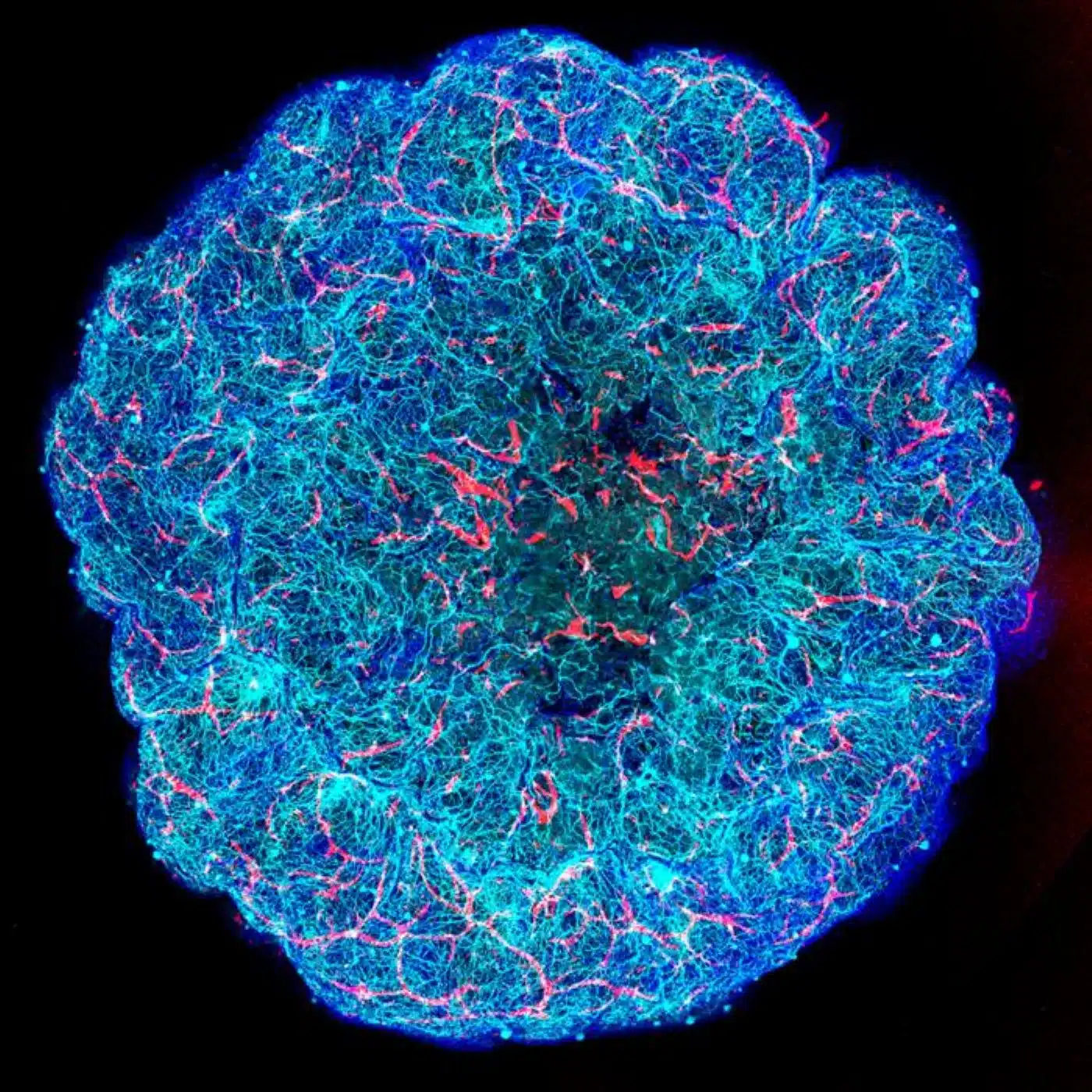
In the parallel study published in Cell Stem Cell, the Blanchard lab utilized their innovative 3D human brain tissue platform, dubbed “miBrains,” to explore how APOE4 promotes the toxic protein accumulation typical of Lewy body dementia, Parkinson’s disease, and Alzheimer’s disease.
Derived from human induced pluripotent stem cells (iPSCs), miBrains faithfully replicate the cellular complexity of living human brain tissue, incorporating neurons, myelinating oligodendrocytes, glial cells, and functional vascular networks.
Using this platform, the researchers uncovered a cascade of metabolic dysfunction:
- Cholesterol Congestion: APOE4 triggers widespread, abnormal cholesterol storage inside astrocytes, the primary metabolic support cells of the central nervous system.
- Lysosomal Breakdown: This lipid accumulation disrupts the astrocytes’ lysosomal machinery, crippling their internal waste-disposal capabilities.
- Pathological Spread: Deprived of effective glial clearance, toxic alpha-synuclein builds up, aggregates, and spreads into neighboring neurons, seeding harmful neurodegenerative inclusions.
These findings identify astrocytic cholesterol trafficking and lysosomal degradation pathways as actionable pharmacological targets to halt toxic protein spread across neurodegenerative diseases.
Scalable, Patient-Specific “miBrains”
A major barrier in neurodegenerative drug discovery has been the difficulty of studying the living human brain before late-stage postmortem damage sets in. The miBrain platform bypasses this limitation by recreating human neurovascular pathology in real time.
“A key advance of our technology is that miBrains with predefined cellular compositions and disease-related factors can be cryopreserved,” says Louise Mesentier-Louro, PhD, Assistant Professor of Neuroscience, and Stem Cell Biology and Regenerative Medicine, and first author of the Cell Stem Cell study. “This capability improves reproducibility and scalability of complex disease modeling and supports more efficient drug development and validation.”
Mount Sinai is currently generating and cryopreserving patient-derived miBrains to create a personalized platform for testing disease vulnerability and drug efficacy, bridging the gap between benchside genetic discovery and clinical therapeutics.
Funding: The Cell study on vascular degeneration in the brain was supported by funds from the National Aeronautics and Space Administration (80ARC022CA004), the National Institute on Aging at the National Institutes of Health (R01AG089533, UH3NS115064, U54AG090669, T32GM146636), The SWT Foundation, and the CureAlz Fund. The Cell Stem Cell study on abnormal protein buildup in the brain was supported by funds from the National Aeronautics and Space Administration (80ARC022CA004), Aligning Science Across Parkinson’s (ASAP-024297) through the Michael J. Fox Foundation for Parkinson’s Research, the National Institute of Neurological Disorders and Stroke and the National Institute on Aging at the National Institutes of Health (R01NS114239, UH3NS115064, 1U54AG090669-01, T32AG04968, F31NS13090), the CureAlz Fund, and The SWT Foundation.
Editorial Notes:
- This article was edited by a Neuroscience News editor.
- Journal paper reviewed in full.
- Additional context added by our staff.
About this Depression Research:
- Media Contact: Stacy Anderson
- Source: Mount Sinai Hospital
- Image Credit: Image credited to The Blanchard Lab/The Mount Sinai Health System
- Original Research is Open Access: Cell (September 24, 2026). “A pericyte-to-myofibroblast transition links APOE4 to cerebrovascular degeneration.” Authors: Braxton R. Schuldt, Dominic Haworth-Staines, Andrea Perez-Arevalo, Diede W.M. Broekaart, Ashley Harlock, Leon Wang, Anna Bright, Georgia Gallagher, Grace Rabinowitz, Alison M. Goate, Towfique Raj, Ana C. Pereira, and Joel W. Blanchard.
- DOI: 10.1016/j.cell.2026.08.058
Abstract
A pericyte-to-myofibroblast transition links APOE4 to cerebrovascular degeneration
Cerebrovascular disease is a major but poorly understood feature of Alzheimer’s disease (AD). The strongest genetic AD risk factor, apolipoprotein E4 (APOE4), is associated with cerebrovascular degeneration, including vascular amyloid deposition and fibrosis.
To uncover how APOE4 promotes cerebrovascular pathology, we assembled a single-cell transcriptomic atlas of human brain vasculature. In APOE4 carriers, pericyte abundance was significantly reduced and accompanied by the emergence of a myofibroblast-like cell population co-expressing contraction and extracellular matrix genes.
Immunostaining confirmed non-vascular myofibroblasts in APOE4 human and mouse brains. We show that APOE4 pericytes transition into myofibroblasts that secrete fibronectin, which promotes vascular amyloid accumulation. Computational and experimental analyses identified elevated transforming growth factor β (TGF-β) signaling as the driver of this pericyte-to-myofibroblast transition.
Inhibition of TGF-β restored pericyte coverage and reduced vascular fibrosis and amyloid to APOE3 levels, revealing a targetable mechanism linking APOE4 to cerebrovascular pathology in AD.


