New research shows that the cells responsible for protecting the brain from infection and inflammation are also responsible for repairing the system of defenses that separates the brain from the rest of the body. These findings have significant clinical implications because certain cardiovascular drugs could possibly impede the brain’s ability to repair itself after a stroke or other injury.
“This study shows that the resident immune cells of the central nervous system play a critical and previously unappreciated role in maintaining the integrity of the blood-brain barrier,” said Maiken Nedergaard, M.D., D.M.Sc., co-director of the Center for Translational Neuromedicine at the University of Rochester Medical Center (URMC) and lead author of the study. “When this barrier is breached it must be rapidly repaired in order to maintain the health of the brain and aid in recovery after an injury – a process that could be impaired by drugs that are intended to prevent this damage in the first place.”
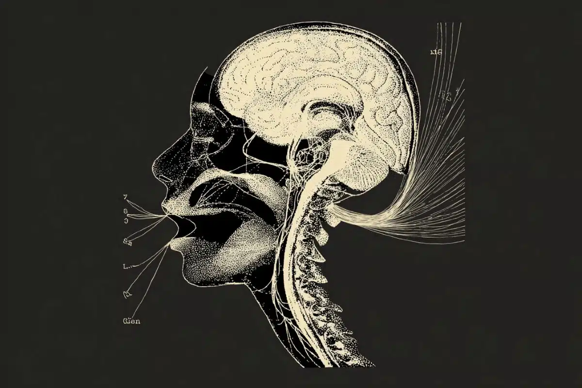
The brain is essentially an independent and separate ecosystem. It possesses a dedicated system of defenses against infection and recently Nedergaard and her colleagues demonstrated that the brain also maintains its own unique process of removing waste. Movement in and out of the brain is tightly controlled through a complex system of gateways and controls that are collectively referred to as the blood-brain barrier (BBB).
When the BBB is breached the brain becomes vulnerable to infection and injury. It is, therefore, imperative that the openings in the BBB are resealed, and quickly. This most frequently occurs during a stroke, which triggers inflammation that can cause the BBB to break down.
The new study, which was published today in the Proceedings of the National Academy of Sciences, reveals that the brain’s immune system, specifically cells called microglia, play a central role in the process of repairing damage to the BBB.
Microglia serve as the brain’s “first responders” and are present throughout the brain and spinal cord. These cells are constantly monitoring their environment, and can be switched on or activated to perform different functions such as control inflammation, destroy pathogens, clean up the debris from dead or damaged cells, and seal off the site of an injury.
Performing experiments in mice, Nedergaard and her colleagues observed that when small holes where made in the BBB, nearby microglia were rapidly mobilized and set about repairing the breach. In most instances, the integrity of the BBB was restored within 10 to 30 minutes.

The team identified a receptor called P2RYX12 that was responsible for activating the microglia and directing them to the site of the damage. This finding is significant because the same receptor is also present on platelets and is one of the targets of blood thinning drugs such as Plavix.
These drugs are given to individuals at risk of heart attack and stroke and helps prevent platelets from binding together to form blood clots that, when they make their way to the brain, can block the flow of blood and trigger a stroke. However, because these drugs also suppress P2RYX12 receptors in microglia, they could potentially impair the ability of the brain to carry out repairs to the BBB once a stroke occurs.
Nedergaard and her team are currently investigating the impact of P2RYX12-blocking drugs on microglia function in the brain.
“Our concern is that while certain types of blood thinning drugs may do a great job preventing strokes, they could have the unintended consequence of making them worse or hindering recovery once they occur,” said Nedergaard.
Additional co-authors of the study include Nanhong Lou, Takahiro Takano, Yong Pei, Anna Xavier, and Steven Goldman with URMC.
Funding: The research was supported by the National Institute of Neurological Disorders and Stroke.
Source: Mark Michaud – University of Rochester Medical Center
Image Source: The image is credited to GerryShaw and is licensed CC BY SA 3.0
Original Research: Abstract for “Purinergic receptor P2RY12-dependent microglial closure of the injured blood–brain barrier” by Nanhong Lou, Takahiro Takano, Yong Pei, Anna L. Xavier, Steven A. Goldman, and Maiken Nedergaard in PNAS. Published online January 11 2016 doi:10.1073/pnas.1520398113
Abstract
Purinergic receptor P2RY12-dependent microglial closure of the injured blood–brain barrier
Microglia are integral functional elements of the central nervous system, but the contribution of these cells to the structural integrity of the neurovascular unit has not hitherto been assessed. We show here that following blood–brain barrier (BBB) breakdown, P2RY12 (purinergic receptor P2Y, G-protein coupled, 12)-mediated chemotaxis of microglia processes is required for the rapid closure of the BBB. Mice treated with the P2RY12 inhibitor clopidogrel, as well as those in which P2RY12 was genetically ablated, exhibited significantly diminished movement of juxtavascular microglial processes and failed to close laser-induced openings of the BBB. Thus, microglial cells play a previously unrecognized protective role in the maintenance of BBB integrity following cerebrovascular damage. Because clopidogrel antagonizes the platelet P2Y12 receptor, it is widely prescribed for patients with coronary artery and cerebrovascular disease. As such, these observations suggest the need for caution in the postincident continuation of P2RY12-targeted platelet inhibition.
“Purinergic receptor P2RY12-dependent microglial closure of the injured blood–brain barrier” by Nanhong Lou, Takahiro Takano, Yong Pei, Anna L. Xavier, Steven A. Goldman, and Maiken Nedergaard in PNAS. Published online January 11 2016 doi:10.1073/pnas.1520398113