While scientists are unsure of the molecular causes of Parkinson’s Disease, they do know the disorder’s tremors and other motor dysfunctions are linked to a loss of dopaminergic neurons located in the substantia nigra, a structure in the midbrain.
Dopaminergic neurons are our main source of dopamine, which is responsible for emotional response and plays an important role in movement. For many years, their loss has been viewed as irreversible.
But a new paper by Brad Morrison’s research group in the Department of Biological Sciences at Boise State University suggests the possibility of regrowth of dopaminergic neurons in adult mammals. Co-authors are Joshua Albright, Iva Stojkovska, Abir Rahman and Connor Brown.
Their results, published in the journal Neuroscience Letters, also show that the rate of replenishment for these neurons is similar to the rate of loss observed in an inflammatory response mouse model of Parkinson’s Disease. This may indicate that inflammatory insult inhibits the natural generation of neurons leading to Parkinson’s Disease.
This is significant because Parkinson’s disease is the most common motor disorder and the second most prevalent neurodegenerative disease. Current medications treat only symptoms and after time lose effectiveness. Learning how to regenerate these neurons could offer potential avenues for preventing neuronal loss and better inform stem cell transplantation efforts.
Morrison hypothesized that cells could regenerate in adults but were somehow being blocked in Parkinson’s Disease, explaining progression of the disease. This goes against current dogma, which posits that only a handful of neuronal populations are able to regenerate in adult mammalian brains.
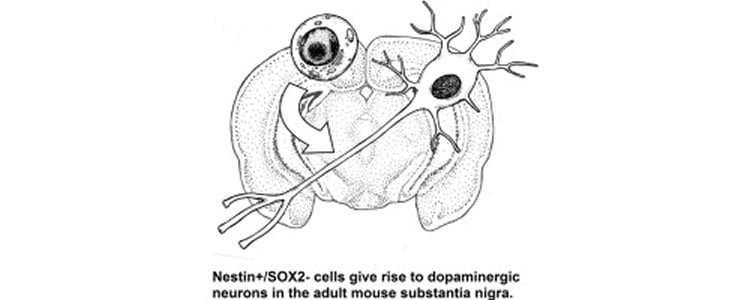
Taking a novel approach, he devised a system to genetically trace the lineage of dopaminergic neurons from stem cells. After removing the gene from stem cells using recombinant DNA technology, he then waited six months. At the end of that time, he saw that removal of the gene affected mature dopaminergic neurons, implying that they must be replenished by stem cells.
He now is characterizing the stem cells responsible for this process as well as looking at the potential correlation of inflammation and a reduction in dopaminergic neurons.
NeuroscienceNews.com would like to thank Brad Morrison for making us aware of this Parkinson’s disease research.
Source: Kathleen Tuck – Boise State University
Image Credit: The image is credited to the researchers/Neuroscience Letters.
Original Research: Full open access research for “Nestin-positive/SOX2−negative cells mediate adult neurogenesis of nigral dopaminergic neurons in mice” by Joshua E. Albright, Iva Stojkovska, Abir A. Rahman, Connor J. Brown, and Brad E. Morrison in Neuroscience Letters. Published online January 20 2016 doi:10.1016/j.neulet.2016.01.019
Abstract
Nestin-positive/SOX2−negative cells mediate adult neurogenesis of nigral dopaminergic neurons in mice
The primary clinical motor symptoms of Parkinson’s disease (PD) result from loss of dopaminergic (DA) neurons in the substantia nigra (SN). Consequently, neurogenesis of this group of neurons in the adult brain has drawn considerable interest for the purpose of harnessing endogenous neurogenerative potential as well as devising better strategies for stem cell therapy for PD. However, the existence of adult neurogenesis for DA neurons within the SN remains controversial. To overcome technical and design limitations associated with previous studies, our group has developed a novel genetic mouse model for assessing adult nigral DA neurogenesis. This system utilizes transgenic mice that express a tamoxifen-activatable Cre recombinase (CreERT2) under the control of the neuronal progenitor cell promoters nestin or Sox2 leading to suppression of the DA neuron marker tyrosine hydroxylase (TH) via excision of exon 1 by flanking loxP sites in adult animals. This study reports that six months following initiation of a six week treatment with tamoxifen mice with nestin-mediated Th excision displayed a significant reduction in TH+ neurons in the SN. This finding indicates that nestin-expressing cells regenerate DA neurons within the SN of adult animals. Interestingly, no reduction was observed in TH+ cells following Sox2-mediated Th excision suggesting that a nestin+/SOX2− precursor cell population drives DA neurogenesis in the adult SN. This information represents a substantial leap in current knowledge of adult DA neurogenesis, will enable improved in vitro and in vivo modeling, as well as facilitate the harnessing of this process for therapeutic intervention for PD.
“Nestin-positive/SOX2−negative cells mediate adult neurogenesis of nigral dopaminergic neurons in mice” by Joshua E. Albright, Iva Stojkovska, Abir A. Rahman, Connor J. Brown, and Brad E. Morrison in Neuroscience Letters. Published online January 20 2016 doi:10.1016/j.neulet.2016.01.019