Summary: In mice genetically engineered to carry human stuttering mutations, vocalization defects are derived from abnormalities in astrocytes in the corpus callosum.
Source: NIH/NIDCD
Researchers believe that stuttering – a potentially lifelong and debilitating speech disorder – stems from problems with the circuits in the brain that control speech, but precisely how and where these problems occur is unknown. Using a mouse model of stuttering, scientists report that a loss of cells in the brain called astrocytes are associated with stuttering. The mice had been engineered with a human gene mutation previously linked to stuttering. The study, which appeared online in the Proceedings of the National Academy of Sciences, offers insights into the neurological deficits associated with stuttering.
The loss of astrocytes, a supporting cell in the brain, was most prominent in the corpus callosum, a part of the brain that bridges the two hemispheres. Previous imaging studies have identified differences in the brains of people who stutter compared to those who do not. Furthermore, some of these studies in people have revealed structural and functional problems in the same brain region as the new mouse study.
The study was led by Dennis Drayna, Ph.D., of the Section on Genetics of Communication Disorders, at the National Institute on Deafness and Other Communication Disorders (NIDCD), part of the National Institutes of Health. Researchers at the Washington University School of Medicine in St. Louis and from NIH’s National Institute of Biomedical Imaging and Bioengineering, and National Institute of Mental Health collaborated on the research.
“The identification of genetic, molecular, and cellular changes that underlie stuttering has led us to understand persistent stuttering as a brain disorder,” said Andrew Griffith, M.D., Ph.D., NIDCD scientific director. “Perhaps even more importantly, pinpointing the brain region and cells that are involved opens opportunities for novel interventions for stuttering – and possibly other speech disorders.”
Stuttering is characterized by pauses and repeated or prolonged sounds, syllables or words, which disrupt the normal flow of speech. People who stutter know what they want to say, but they have trouble saying it. The condition is most commonly seen in young children who typically outgrow the problem. However, for 1 in 4 children who experience early stuttering, the condition persists as a lifelong communication problem. It is estimated that as many as 1% of adults in the United States are affected by stuttering.
“The brain imaging studies of people who stutter are important, but those results can only take us so far,” said Drayna. One challenge, he said, is that the imaging studies cannot decipher if the differences contribute to stuttering or are an effect of stuttering.
“By taking a genetic approach, we have been able to begin deciphering the neuropathology of stuttering, first at the molecular level by identifying genetic mutations, and now at the cellular level,” added Drayna.
Earlier research by Drayna and colleagues has identified several genes associated with stuttering. In this study, the researchers set out to identify changes in the brain brought on by the mutations in a gene called GNPTAB, one of the genes previously linked to stuttering. The scientists engineered this human stuttering mutation into the mice to create a mouse model. The mice with the GNPTAB mutation had long pauses in their stream of vocalizations, similar to those found in people with the same mutation. Like people who stutter, the mice were normal in all other ways, reinforcing earlier research that suggests that the mice can serve as a valid animal model for important features of this disorder.
The investigators next examined brain tissue from the mice and found a decrease in astrocytes, but not other cell types, in the animals with the genetic mutation compared to the mice without the mutation. Astrocytes play a critical role in supporting nerve cells by carrying out a wide range of functions, such as supplying nerve cells with oxygen and nutrients and providing structural support.
The loss of astrocytes was more pronounced in the corpus callosum of the mutant mice. In addition, using advanced magnetic resonance imaging (MRI) methods, the researchers detected reduced local volume of the corpus callosum in the mutant mice despite normal diffusion tensor MRI values, providing further support for a defect in this brain region.

Containing as many as 200 million nerve fibers, the corpus callosum enables communication between the brain’s left and right hemispheres, helping to integrate signals for processes that involve both hemispheres, such as physical coordination and use of language.
Follow-up experiments in which the GNPTAB human stuttering mutation was introduced into individual brain cell types–rather than the entire mouse–confirmed that the vocalization defect is specific to astrocytes. The mice did not have the stuttering-like vocalizations when the mutation was engineered into other types of brain cells.
All of the stuttering genes that have been identified over the past decade are involved in intracellular trafficking, the process that cells use to move proteins and other components to their correct locations within the cell. Defects in cellular trafficking have been linked to other neurological disorders, such as amyotrophic lateral sclerosis (ALS), Parkinson’s disease, and Alzheimer’s disease, suggesting that certain nerve cell pathways are particularly sensitive to impairment of this process. The research does not indicate, however, that persistent stuttering is an early indicator of these other disorders.
If future research confirms that stuttering in people with GNPTAB mutations derives from a loss of astrocytes in the brain, these findings could open the door to new therapeutic strategies for some people with persistent developmental stuttering by targeting associated molecular pathways and cells.
Funding: This work was supported by the Intramural Research Program of the NIH, by the NIDCD under intramural Grant Z1A-000046-18, the National Institute of Mental Health Rodent Behavioral Core, and the National Heart, Lung, and Blood Institute Animal MRI Core.
Source:
NIH/NIDCD
Media Contacts:
NIDCD News – NIH/NIDCD
Image Source:
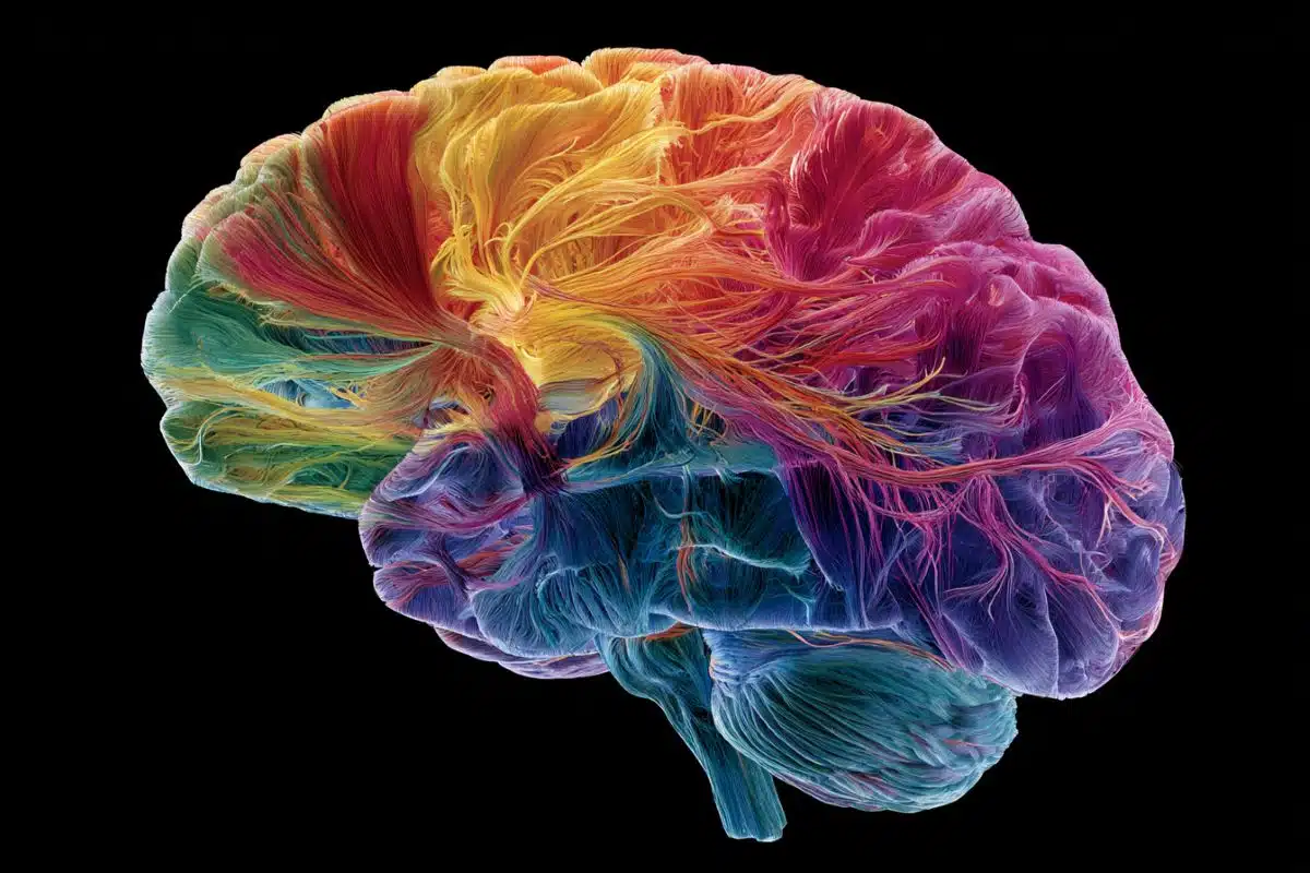
The image is credited to Tae-Un Han, Ph.D., National Institutes on Deafness and Other Communication Disorders.
Original Research: Closed access
“Human GNPTAB stuttering mutations engineered into mice cause vocalization deficits and astrocyte pathology in the corpus callosum”. Tae-Un Han, Jessica Root, Laura D. Reyes, Elizabeth B. Huchinson, Johann du Hoffmann, Wang-Sik Lee, Terra D. Barnes, and Dennis Drayna.
PNAS. doi:10.1073/pnas.1901480116
Abstract
Human GNPTAB stuttering mutations engineered into mice cause vocalization deficits and astrocyte pathology in the corpus callosum
Stuttering is a common neurodevelopmental disorder that has been associated with mutations in genes involved in intracellular trafficking. However, the cellular mechanisms leading to stuttering remain unknown. Engineering a mutation in N-acetylglucosamine-1-phosphate transferase subunits α and β (GNPTAB) found in humans who stutter into the mouse Gnptab gene resulted in deficits in the flow of ultrasonic vocalizations similar to speech deficits of humans who stutter. Here we show that other human stuttering mutations introduced into this mouse gene, Gnptab Ser321Gly and Ala455Ser, produce the same vocalization deficit in 8-day-old pup isolation calls and do not affect other nonvocal behaviors. Immunohistochemistry showed a marked decrease in staining of astrocytes, particularly in the corpus callosum of the Gnptab Ser321Gly homozygote mice compared to wild-type littermates, while the staining of cerebellar Purkinje cells, oligodendrocytes, microglial cells, and dopaminergic neurons was not significantly different. Diffusion tensor imaging also detected deficits in the corpus callosum of the Gnptab Ser321Gly mice. Using a range of cell type-specific Cre-drivers and a Gnptab conditional knockout line, we found that only astrocyte-specific Gnptab-deficient mice displayed a similar vocalization deficit. These data suggest that vocalization defects in mice carrying human stuttering mutations in Gnptab derive from abnormalities in astrocytes, particularly in the corpus callosum, and provide support for hypotheses that focus on deficits in interhemispheric communication in stuttering.