

Summary:
Biomedical engineers and developmental biologists at Columbia University have engineered 3D human stem cell models that recreate the earliest, previously inaccessible stages of human placental formation. Published in Nature Cell Biology, the research led by Dr. Mijo Simunovic reveals how human pluripotent stem cells, temporarily reset to a naive state, can be biochemically steered to suppress Hippo signaling, initiating the embryo’s first major cell-fate decision.
The cells self-organize over three weeks into complex chorionic tissue architectures with fluid-filled lacunae that mirror early implantation, unlocking a mechanistic platform to investigate early pregnancy failure, preeclampsia, and fetal growth restriction.
Key Facts:
- Modeling the First Cell Fate: The 3D stem-cell platform captures the developmental window where embryonic cells first diverge to create the chorion, the structural foundation of the human placenta, overcoming ethical and optical barriers of in vivo uterine implantation.
- The Hippo Signaling Trigger: By briefly resetting human pluripotent stem cells to naive pluripotency and selectively downregulating the mechanosensitive Hippo signaling pathway, researchers activated the native human placental program without needing external tissue scaffolds.
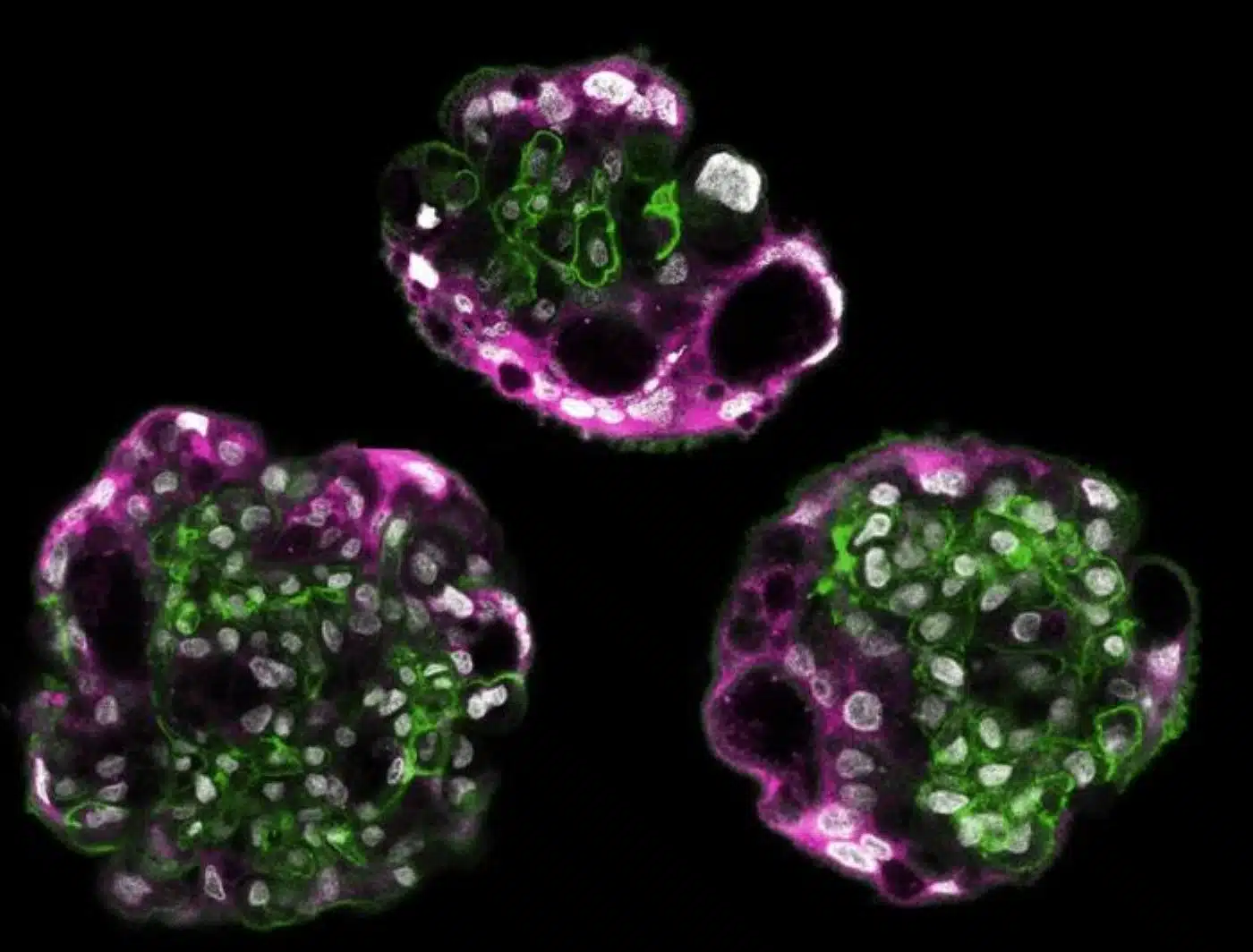
- Spontaneous 3D Self-Organization: Over a three-week culture window, the cells autonomously differentiated into the major cell types of the chorion and self-assembled into architectural layers complete with fluid-filled lacunae that mimic spaces where maternal blood vessels normally connect.
Source: Columbia University / Nature Cell Biology
The placenta is the transient, life-sustaining organ that anchors mammalian pregnancy, acting as the fetus’s lungs, kidneys, liver, and immune barrier. Despite its fundamental role in human reproduction, the earliest days of placental formation have remained an impenetrable biological black box.
Because early implantation occurs deep within the human endometrium shortly after conception, it cannot be optically tracked or ethically biopsied in vivo. Furthermore, comparative animal studies offer limited insight; the molecular and physical dynamics of human blastocyst implantation diverge sharply from those of standard laboratory rodent models.
To overcome this longstanding barrier, a team led by Dr. Mijo Simunovic at Columbia University has developed a functional three-dimensional, lab-grown stem cell model that successfully recapitulates the earliest stages of human placenta formation.
Published in Nature Cell Biology, the work provides an experimental window into how pregnancy begins and why it so frequently terminates in early miscarriage.
Recreating the Embryo’s First Identity Choice
Within days of fertilization, the human blastocyst faces its very first developmental crossroad. Cells on the outer perimeter must separate from the inner cell mass (which forms the embryo proper) and commit to an extraembryonic fate.
These outer cells establish the chorion, the foundational tissue that invades maternal uterine tissue to assemble the functional placenta. Molecular errors during this crucial transition can cause implantation failure, recurrent pregnancy loss, or devastating mid-to-late gestational pathologies, including preeclampsia and intrauterine fetal growth restriction (FGR).
To model this transition in vitro, Simunovic’s laboratory, bridging Columbia’s Department of Chemical Engineering and Department of Genetics and Development, focused on the Hippo signaling pathway. Known primarily for regulating organ size, contact inhibition, and mechanotransduction, Hippo signaling acts as the primary molecular compass governing this initial divergence.
The researchers initiated their platform using human pluripotent stem cells:
- Naive Reversion: The stem cells were transiently reset into a naive pluripotent state, opening an expanded window of developmental plasticity.
- Hippo Modulation: The team introduced targeted biochemical cues to downregulate Hippo signaling, matching the exact mechanochemical conditions seen in the outer blastocyst.
- Autonomous Morphogenesis: Once this regulatory switch flipped, the cells executed their native extraembryonic genetic blueprint without requiring step-by-step external direction.
“What is remarkable is that we don’t have to tell each cell what to become or where to go,” says Simunovic. “Once we set the developmental process in motion, the cells begin to organize themselves into a complex tissue. We can now study how that happens.”
Formation of 3D Layers and Placental Lacunae
Over a 21-day timeline, the stem cell cultures transitioned from flat cultures into complex 3D organotypic structures.
The cells differentiated into the principal lineages of the early human chorion and spontaneously migrated into anatomical layers that closely resemble in vivo placental architecture.
Remarkably, the 3D structures formed internal, fluid-filled cavities identical to placental lacunae—specialized microscopic chambers that emerge during human implantation to capture and interface with invading maternal spiral arteries.
“Being able to watch a human placenta form in the lab should be a boon to both basic embryology and clinical research,” explains Simunovic. “We hope it will be useful in studying how the placenta forms and how its formation can go awry, which is one of the leading causes of early pregnancy failure.”
New Horizons for Reproductive Medicine
Because human implantation is distinct from other mammals, this platform enables researchers to test human-specific hypotheses regarding mechanical forces, endocrine signals, and cellular cross-talk at the maternal-fetal boundary.
Future iterations of the model will co-culture these placental structures with endometrial organoids, allowing scientists to simulate the complete implantation sequence in vitro. By identifying the molecular checkpoints where placental formation fails, the system provides a preclinical engine to uncover early diagnostic biomarkers and targeted therapeutics to treat infertility, recurrent miscarriage, and pregnancy-induced hypertension.
Questions and Answers
Why has early human placenta formation been so difficult to study?
Early placental formation occurs deep inside the uterus days after fertilization, making direct observation impossible without terminating pregnancy. Furthermore, human embryo implantation is biologically and architecturally distinct from rodents, rendering traditional animal models inadequate for studying human-specific defects.
How did the Columbia researchers trigger placental differentiation from stem cells?
The researchers temporarily reset human pluripotent stem cells to a naive state of expanded potency and then applied targeted biochemical signals to suppress the Hippo signaling pathway. This suppression mirrored the natural developmental switch that directs the embryo’s outer cells to form the chorion.
What structures did the lab-grown placental models develop?
Over three weeks, the cells spontaneously differentiated into the major chorionic lineages and organized into distinct 3D layers. They also developed fluid-filled cavities resembling placental lacunae, the early anatomical structures that connect with maternal blood vessels during uterine implantation.
Editorial Notes:
- This article was edited by a Neuroscience News editor.
- Journal paper reviewed in full.
- Additional context added by our staff.
About this genetics Research:
- Media Contact: Helen Garey
- Source: Columbia University
- Image Credit: Image credited to Mijo Simunovic, Columbia University
- Original Research is Open Access: Nature Cell Biology (Nov 3, 2026). “Reconstructing the emergence of the human chorion via Hippo-mediated trophoblast induction.” Authors: Miaoci Zhang, Ryan L Lim, Alice H. Reis, Walter Piszker, Wallis W. Boyd, Athena Pagon, Aman Mahajan, Linlin Wu, Cheng Zhao, Sophie Petropoulos, Carlotta Ronda & Mijo Simunovic.
- DOI: 10.1038/s41556-026-02082-9
Abstract
Reconstructing the emergence of the human chorion via Hippo-mediated trophoblast induction
Differential Hippo–LATS–YAP signalling controls the first mammalian cell-fate decision, yet how human trophectoderm specification progresses to chorion formation remains unknown. Here we establish a developmentally informed model in which LATS kinase inhibition initiates the trophectoderm trajectory without exogenous morphogens.
Stage matching to an integrated embryo–chorion single-cell reference revealed ordered progression from early trophectoderm to secondary chorionic lineages. Whole-genome bisulfite sequencing showed a naive-like hypomethylated state followed by remethylation, with key known placenta-associated imprinted domains retaining methylation and allelic bias. We then established a three-dimensional human chorion model by agitating induced-cell aggregates in suspension, yielding secondary chorionic lineages that self-organized with outward-facing trophoblast polarity and inside-out stromal–epithelial topology.
Endogenous WNT signalling was dispensable for initial trophectoderm induction but regulated later chorionic lineage balance. Isolating CLDN6+ITGA6− cells identified a proEVT-like intermediate with villous and extravillous potential.
Together, this framework connects human trophectoderm induction to three-dimensional chorion modelling and enables investigation of placental specification.


