Summary: Researchers developed a stem cell-derived model of the human embryo in the lab. This innovative model, created from pluripotent stem cells, enables experimental modeling of embryonic development during the second week of pregnancy, a critical period often associated with pregnancy loss.
The research aims to enhance our understanding of genetic disorders, early pregnancy loss, and the origins of organs and specialized cells. This new advance may reduce the reliance on donated human embryos in research.
Key Facts:
- Cambridge scientists have created a model of the human embryo in the lab using reprogrammed human stem cells.
- The model allows for experimental modeling of embryonic development during the second week of pregnancy, a time when many pregnancies fail.
- This breakthrough could enhance our understanding of genetic disorders, early pregnancy loss, and the developmental origins of organs and specialized cells, and could reduce the need for donated human embryos in research.
Source: University of Cambridge
Cambridge scientists have created a stem cell-derived model of the human embryo in the lab by reprogramming human stem cells. The breakthrough could help research into genetic disorders and in understanding why and how pregnancies fail.
Published today in the journal Nature, this embryo model is an organized three-dimensional structure derived from pluripotent stem cells that replicate some developmental processes that occur in early human embryos.
Use of such models allows experimental modeling of embryonic development during the second week of pregnancy. They can help researchers gain basic knowledge of the developmental origins of organs and specialised cells such as sperm and eggs, and facilitate understanding of early pregnancy loss.

“Our human embryo-like model, created entirely from human stem cells, gives us access to the developing structure at a stage that is normally hidden from us due to the implantation of the tiny embryo into the mother’s womb,” said Professor Magdalena Zernicka-Goetz in the University of Cambridge’s Department of Physiology, Development and Neuroscience, who led the work.
She added: “This exciting development allows us to manipulate genes to understand their developmental roles in a model system. This will let us test the function of specific factors, which is difficult to do in the natural embryo.”
In natural human development, the second week of development is an important time when the embryo implants into the uterus. This is the time when many pregnancies are lost.
The new advance enables scientists to peer into the mysterious ‘black box’ period of human development – usually following implantation of the embryo in the uterus – to observe processes never directly observed before.
Understanding these early developmental processes holds the potential to reveal some of the causes of human birth defects and diseases, and to develop tests for these in pregnant women.
Until now, the processes could only be observed in animal models, using cells from zebrafish and mice, for example.
Legal restrictions in the UK currently prevent the culture of natural human embryos in the lab beyond day 14 of development: this time limit was set to correspond to the stage where the embryo can no longer form a twin.
Until now, scientists have only been able to study this period of human development using donated human embryos. This advance could reduce the need for donated human embryos in research.
Zernicka-Goetz says the while these models can mimic aspects of the development of human embryos, they cannot and will not develop to the equivalent of postnatal stage humans.
Over the past decade, Zernicka-Goetz’s group in Cambridge has been studying the earliest stages of pregnancy, in order to understand why some pregnancies fail and some succeed.
In 2021 and then in 2022 her team announced in Developmental Cell, Nature and Cell Stem Cell journals that they had finally created model embryos from mouse stem cells that can develop to form a brain-like structure, a beating heart, and the foundations of all other organs of the body.
The new models derived from human stem cells do not have a brain or beating heart, but they include cells that would typically go on to form the embryo, placenta and yolk sac, and develop to form the precursors of germ cells (that will form sperm and eggs).
Many pregnancies fail at the point when these three types of cells orchestrate implantation into the uterus begin to send mechanical and chemical signals to each other, which tell the embryo how to develop properly.
There are clear regulations governing stem cell-based models of human embryos and all researchers doing embryo modelling work must first be approved by ethics committees. Journals require proof of this ethics review before they accept scientific papers for publication. Zernicka-Goetz’s laboratory holds these approvals.
“It is against the law and FDA regulations to transfer any embryo-like models into a woman for reproductive aims. These are highly manipulated human cells and their attempted reproductive use would be extremely dangerous,” said Dr Insoo Hyun, Director of the Center for Life Sciences and Public Learning at Boston’s Museum of Science and a member of Harvard Medical School’s Center for Bioethics.
Zernicka-Goetz also holds position at the California Institute of Technology and is NOMIS Distinguished Scientist and Scholar Awardee.
Funding: The research was funded by the Wellcome Trust and Open Philanthropy.
About this stem cell and neurodevelopment research news
Author: Craig Brierley
Source: University of Cambridge
Contact: Craig Brierley – University of Cambridge
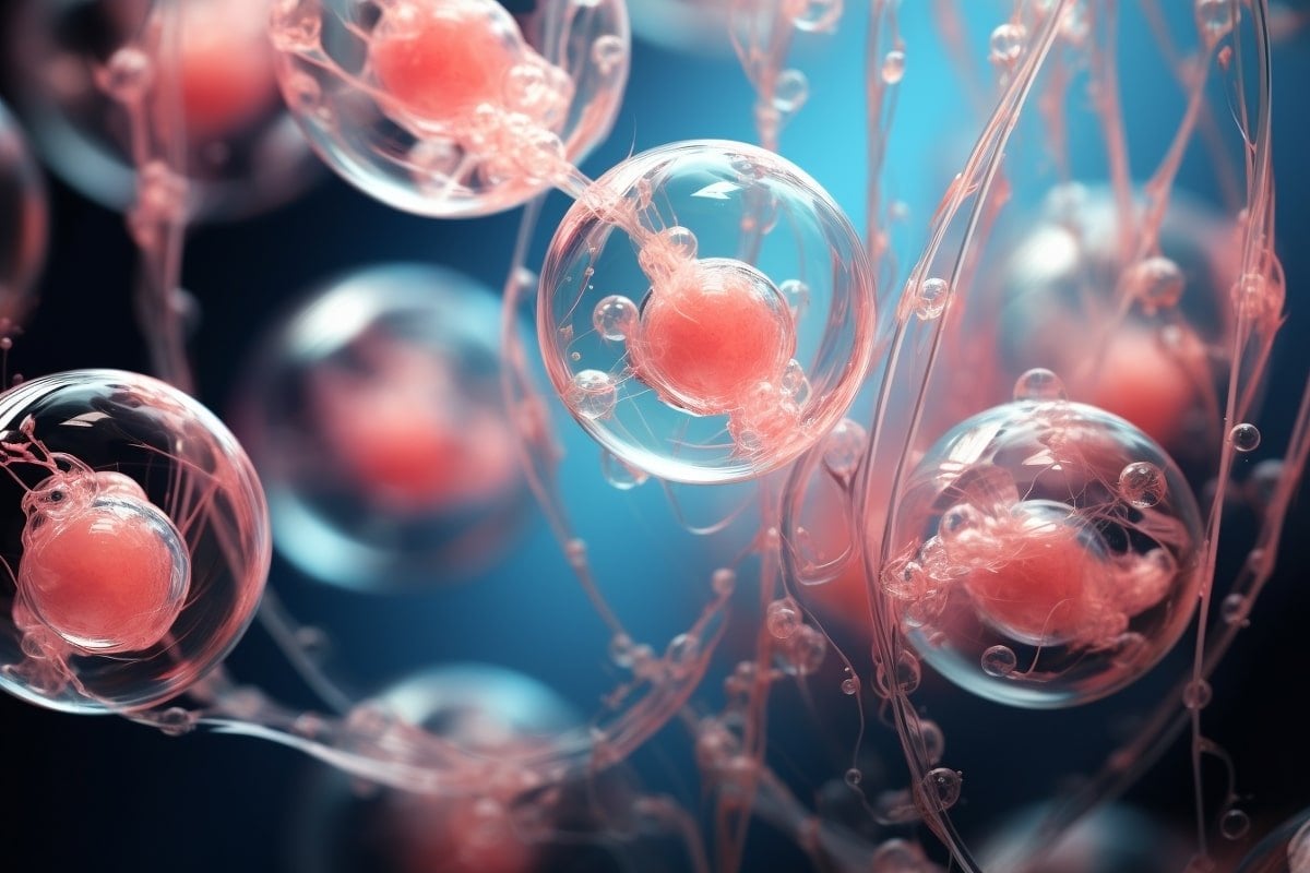
Image: The image is credited to Neuroscience News
Original Research: Closed access.
“A model of the post-implantation human embryo derived from pluripotent stem cells” by Magdalena Zernicka-Goetz et al. Nature
Abstract
A model of the post-implantation human embryo derived from pluripotent stem cells
The human embryo undergoes morphogenetic transformations following implantation into the uterus, yet our knowledge of this crucial stage is limited by the inability to observe the embryo in vivo. Stem cell-derived models of the embryo are important tools to interrogate developmental events and tissue-tissue crosstalk during these stages.
Here, we establish a model of the human post-implantation embryo, a human embryoid, comprised of embryonic and extraembryonic tissues.
We combine two types of extraembryonic-like cells generated by transcription factor overexpression with wildtype embryonic stem cells and promote their self-organization into structures that mimic several aspects of the post-implantation human embryo. These self-organized aggregates contain a pluripotent epiblast-like domain surrounded by extraembryonic-like tissues.
Our functional studies demonstrate that the epiblast-like domain robustly differentiates to amnion, extraembryonic mesenchyme, and primordial germ cell-like cells in response to BMP cues. In addition, we identify an inhibitory role for SOX17 in the specification of anterior hypoblast-like cells.
Modulation of the subpopulations in the hypoblast-like compartment demonstrated that extraembryonic-like cells impact epiblast-like domain differentiation, highlighting functional tissue-tissue crosstalk.
In conclusion, we present a modular, tractable, integrated model of the human embryo that will allow us to probe key questions of human post-implantation development, a critical window when significant numbers of pregnancies fail.


