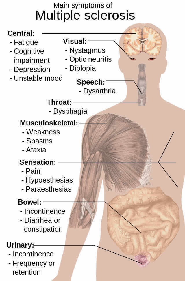
Summary: Researchers report spinal cord cross sectional area is not a good predictor of axonal loss in multiple sclerosis.
Source: Queen Mary University of London.
Researchers from Queen Mary University of London have now sampled spinal cords of 13 people with MS and five healthy controls, and found that spinal cord cross sectional area is not a good predictor of axonal loss
It is commonly thought that in MS, the loss of axons (nerve fibres) contributes to the chronic disability found in many patients. This has led to the wide use of MRI to measure the cross sectional area of the spinal cord in order to predict disability.
But researchers from Queen Mary University of London have now sampled spinal cords of thirteen people with MS and five healthy controls, and found that spinal cord cross sectional area is not a good predictor of axonal loss.

Lead researcher Klaus Schmierer said: “The lack of association between axonal loss and spinal cord cross sectional area significantly changes our understanding of chronic disability in MS.
“The nature of the spinal cord as a highly organised and largely autonomous network needs to be appreciated. We need to identify other factors which – over and above axonal loss – determine the collapse of the spinal cord network and lead to the functional deficits seen in MS.
“In spinal cord trauma, people with less than 10% of their spinal cord axons may still be able to have useful lower limb movement, but in MS, patients with as much as 40% of their axons retained, as shown in our study, are almost invariably wheelchair bound. So there is clearly something happening here which we’ve yet to understand.”
The researchers say that finding other factors that cause the chronic disability seen in MS could help identify targets for new treatments.
The team’s preliminary results indicate that the loss of synaptic connections in the MS spinal cord is substantial, and that this could be the missing link that is driving disability.
Source: Joel Winston – Queen Mary University of London
Image Source: NeuroscienceNews.com image is in the public domain.
Original Research: Full open access research for “Axonal loss in the multiple sclerosis spinal cord revisited” by Natalia Petrova, Daniele Carassiti, Daniel R. Altmann, David Baker, and Klaus Schmiererin Brain Pathology. Published online May 7 2017 doi:10.1111/bpa.12516
[cbtabs][cbtab title=”MLA”]Queen Mary University of London”Loss of Spinal Nerve Fibers Not the Only Cause of Disability in MS.” NeuroscienceNews. NeuroscienceNews, 10 May 2017.
<https://neurosciencenews.com/spinal-neurons-axons-ms-6638/>.[/cbtab][cbtab title=”APA”]Queen Mary University of London(2017, May 10). Loss of Spinal Nerve Fibers Not the Only Cause of Disability in MS. NeuroscienceNew. Retrieved May 10, 2017 from https://neurosciencenews.com/spinal-neurons-axons-ms-6638/[/cbtab][cbtab title=”Chicago”]Queen Mary University of London”Loss of Spinal Nerve Fibers Not the Only Cause of Disability in MS.” https://neurosciencenews.com/spinal-neurons-axons-ms-6638/ (accessed May 10, 2017).[/cbtab][/cbtabs]
Abstract
Axonal loss in the multiple sclerosis spinal cord revisited
Preventing chronic disease deterioration is an unmet need in people with multiple sclerosis, where axonal loss is considered a key substrate of disability. Clinically, chronic multiple sclerosis often presents as progressive myelopathy. Spinal cord cross-sectional area (CSA) assessed using MRI predicts increasing disability and has, by inference, been proposed as an indirect index of axonal degeneration. However, the association between CSA and axonal loss, and their correlation with demyelination, have never been systematically investigated using human post mortem tissue. We extensively sampled spinal cords of seven women and six men with multiple sclerosis (mean disease duration= 29 years) and five healthy controls to quantify axonal density and its association with demyelination and CSA. 396 tissue blocks were embedded in paraffin and immuno-stained for myelin basic protein and phosphorylated neurofilaments. Measurements included total CSA, areas of (i) lateral cortico-spinal tracts, (ii) gray matter, (iii) white matter, (iv) demyelination, and the number of axons within the lateral cortico-spinal tracts. Linear mixed models were used to analyze relationships. In multiple sclerosis CSA reduction at cervical, thoracic and lumbar levels ranged between 19 and 24% with white (19–24%) and gray (17–21%) matter atrophy contributing equally across levels. Axonal density in multiple sclerosis was lower by 57–62% across all levels and affected all fibers regardless of diameter. Demyelination affected 24–48% of the gray matter, most extensively at the thoracic level, and 11–13% of the white matter, with no significant differences across levels. Disease duration was associated with reduced axonal density, however not with any area index. Significant association was detected between focal demyelination and decreased axonal density. In conclusion, over nearly 30 years multiple sclerosis reduces axonal density by 60% throughout the spinal cord. Spinal cord cross sectional area, reduced by about 20%, appears to be a poor predictor of axonal density.
“Axonal loss in the multiple sclerosis spinal cord revisited” by Natalia Petrova, Daniele Carassiti, Daniel R. Altmann, David Baker, and Klaus Schmiererin Brain Pathology. Published online May 7 2017 doi:10.1111/bpa.12516


