

Summary: Researchers developed two innovative high-throughput single-cell genomic tools, IRISeq and EnrichSci, to decode the molecular mysteries of aging.
These technologies allow scientists to examine the molecular states of tens of millions of cells simultaneously, revealing how cellular neighborhoods and rare cell populations drive age-related decline.
Key Research Findings
- Optics-Free Spatial Mapping (IRISeq): This tool uses DNA as a “molecular ruler” to map tissue layouts without a microscope. Millions of barcoded beads capture gene expression and exchange signals with neighbors to reconstruct cellular locations.
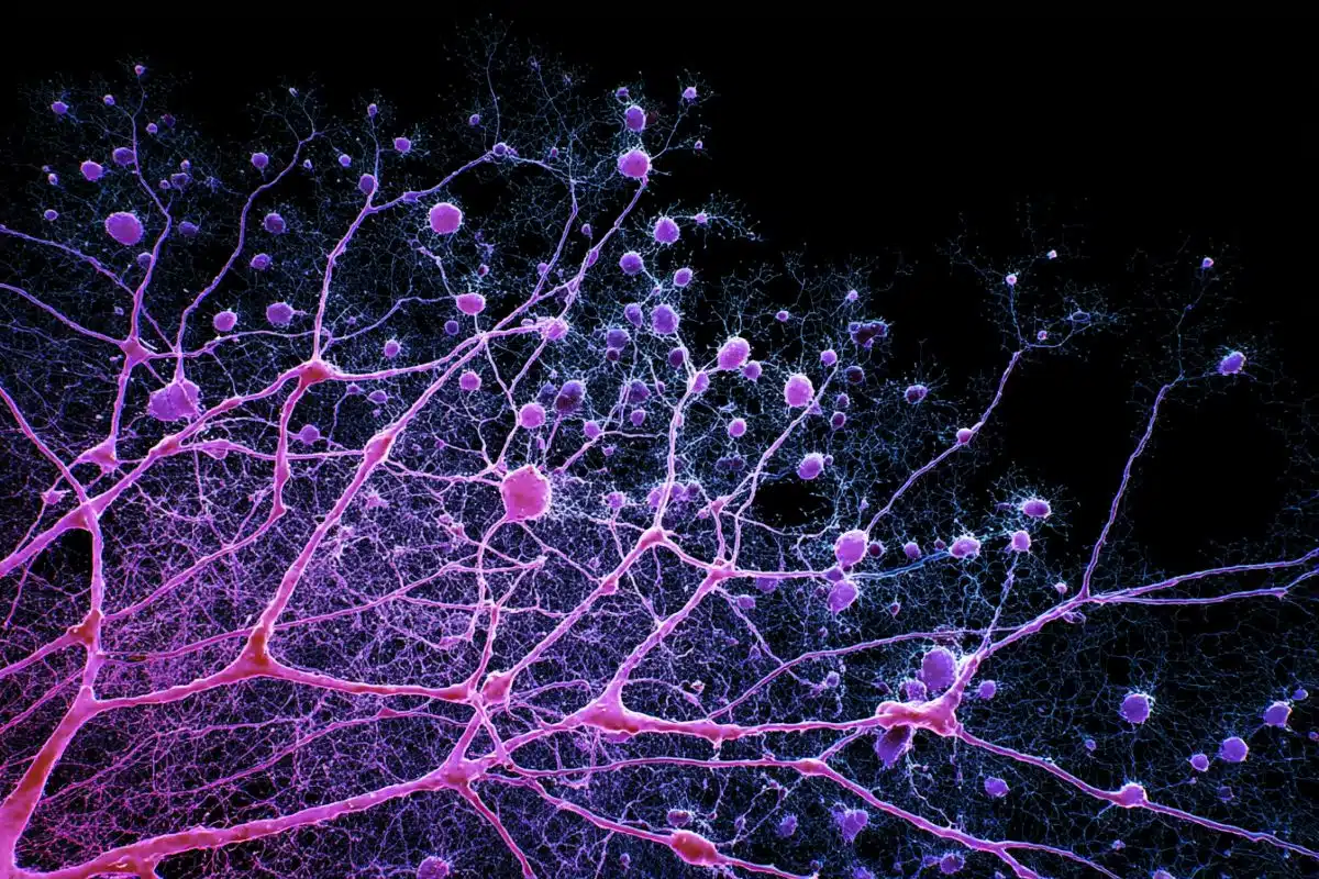
- Inflammatory Neighborhoods: Using IRISeq, researchers discovered clusters of inflammatory microglia, oligodendrocytes, and astrocytes in the brain’s white matter. These findings suggest white matter is a highly vulnerable region where disease-associated states reinforce each other.
- Ventricle-Specific Inflammation: The team found that lymphocytes drive inflammation specifically near the brain’s ventricles (fluid-filled spaces), a localized activity that traditional methods would have missed.
- Targeting Rare Cells (EnrichSci): This method isolates and enriches rare, biologically relevant cell populations, such as specific oligodendrocyte subtypes, before performing deep molecular analysis.
- Unexpected Genetic Changes: Researchers found that while many genes maintain stable expression levels during aging, their exons (parts of genes that form mature RNA) undergo significant changes related to alternative splicing.
Source: Rockefeller University
While much is mysterious about the aging process, change over time remains its cornerstone. The biological shifts that accompany aging seemingly occur in many cells in the body. The problem is, we have tens of billions of cells, and what the changes may be in most of those cells remains unknown, in part due to traditional technical limitations.
That’s why Rockefeller’s Junyue Cao has developed a suite of high-throughput single-cell genomic analysis tools that can examine how aging affects the molecular state of tens of millions of cells in the brain simultaneously.
Now Cao’s Laboratory of Single-Cell Genomics and Population Dynamics has produced two new tools and concepts for studying the molecular changes, gene expression, and intercellular dynamics of aging.
In papers published in Nature Neuroscience and Cell Genomics, Cao and his team describe each of the new technologies as well as the biological insights they have already yielded.
“These approaches essentially take two different paths to understanding different elements of cellular dynamics and the changing molecular processes that accompany aging,” says Cao.
Molecular neighbors
Cao’s lab specializes in developing new techniques of high-throughput single-cell sequencing, which reveals the genetic expression and molecular dynamics of millions of individual cells simultaneously.
While these methods can be used for various analyses, Cao’s lab focuses on aging. His team has previously used single-cell sequencing to identify rare brain cell types, track how brain cells age, pinpoint the cells most vulnerable to age-related decline, and discover that aging may be a developmental stage triggered by specific molecular cues.
The two new approaches Cao’s lab has developed, IRISeq and EnrichSci, tackle cellular aging from different angles.
IRISeq capitalizes on recent discoveries showing that DNA can act like a kind of molecular barcode or even a ruler, recording which molecules are close to one another, says Abdulraouf Abdul, an M.D.-Ph.D. student in Cao’s lab. “For centuries, scientists have relied on microscopes to study tissues and understand how cells are organized. We wondered, could DNA itself be used to map entire tissues without using a microscope at all?”
The technique was designed to do just that. In work led by Abdul and Weirong Jiang, a research associate in the Cao lab, the team developed an optics-free, high-throughput approach that uses millions of barcoded, micrometer-sized beads to capture local gene expression information across tissue. The beads exchange DNA-based signals with nearby beads, allowing researchers to piece together where cells are located in the tissue—and all without a microscope.
Abdul and his colleagues described the work in Nature Neuroscience.
“With IRISeq, we can rebuild the layout of tissues at different levels of detail—almost like zooming in and out on a map—without ever taking a single picture. That means we can study very large pieces of tissues or many tissue sections in a way that would be much harder or more expensive to do with traditional imaging methods,” he says. “In essence, we turned sequencing into a new way of ‘seeing’ biology at a fraction of the cost.”
Adds Cao, “We can use this map interaction to see how the cells are driven by their external cell interactions during the aging process. It can probe the interactions of virtually any two—or more—types of cells anywhere in the brain at the same time.”
Using this approach, the team mapped inflammatory cellular neighborhoods in the aging brain. They found that inflammatory subtypes of microglia, oligodendrocytes, and astrocytes tend to cluster together in white matter and interact with one another. These findings suggest that white matter may be a particularly vulnerable region of the aging brain where disease-associated cellular states emerge and reinforce each other.
For example, they found that immune cells called lymphocytes play a major role in driving inflammation in the aging brain in a very specific way.
“Their activity is concentrated in certain regions, especially near the brain’s fluid-filled spaces known as ventricles,” Abdul says. “Without spatial information, this kind of localized immune activity would have been easy to miss.”
“Knowing both the types of cells that cluster together and where they do so can provide potential targets for anti-aging interventions,” Cao says.
Changes in unexpected places
The second, called EnrichSci, published in Cell Genomics, is a single-nucleus RNA sequencing method that first targets and isolates rare but biologically relevant cells in a mixed population of cells, elevating the percentage of the target cell type in the sample. After enriching for the rare target cells, EnrichSci then zooms in on each cell’s molecular programming.
The researchers applied EnrichSci in the aging mouse brain to enrich for rare cell populations they’d previously identified as especially prone to problematic shifts during aging, among them subtypes of oligodendrocytes, which are found exclusively in the central nervous system.
These cells ensheath neuronal axons in the brain and spinal cord and are linked to neurodegenerative diseases. In these aging subtypes the researchers uncovered changes in both gene expression and in influential genetic elements called exons, which are key to the post-transcriptional regulation of genes.
“Exons are the parts of genes that make up the mature RNA transcripts that are either translated into proteins or serve other biological functions,” describes first author Andrew Liao, an M.D.-Ph.D student in Cao’s lab.
The exonic changes they identified revealed that post-transcriptional regulation plays an important role in how oligodendrocytes age and could offer new targets for modulating these changes in age-related neurodegeneration.
“Surprisingly, we also found that many genes don’t undergo significant changes in expression during the aging process, but their exons do,” adds Cao. “These changes were related to alternate splicing, a key mechanism for creating different protein functions. But such changes can also be linked to many diseases, including cancer.”
Beyond aging
The researchers hope their techniques function as both clinical and research tools for diagnosing disease and uncovering new biology across a wide range of conditions.
“We are already scaling IRISeq to study aging and pharmacological interventions at a scale that was previously unfeasible. At the heart of this vision is the idea that cells don’t act in isolation—their behavior depends on where they are and which other cells surround them,” Abdul says.
“Studying cells without that context is like reading individual words from a book after the pages have been torn apart. By preserving spatial relationships between cells, IRISeq enables the study of how tissues function, change, and respond to disease across larger sample sets and broader contexts.”
Liao hopes to expand EnrichSci to jointly profile both RNA and chromatin accessibility. “Such a co-assay would be able to capture gene and exon expression changes as well as their underlying epigenetic changes,” he says.
“We hope to apply this method to further study the aging changes in oligodendrocytes and other vulnerable cell types in age-related neurodegeneration.”
Cao says their new techniques can be used to study cellular dynamics in many biological contexts. “While my lab focuses on the cell population dynamics associated with aging, these techniques can be used for any disease model system. For example, IRISeq can be used to study immune cell interactions during the cancer progression process, and EnrichSci can illuminate post-transcriptional changes that might be involved in disease progression.”
Key Questions Answered:
A: IRISeq turns DNA into a sensor. By using microscopic beads that “talk” to each other via DNA signals, researchers can reconstruct exactly where every cell was located based on which beads were neighbors, essentially “calculating” the map rather than photographing it.
A: It was previously thought that aging meant genes simply turned “on” or “off”. This research shows that genes can stay “on,” but the way they are put together (via exons) changes. This “alternative splicing” can create different protein functions, potentially contributing to neurodegeneration or cancer.
A: IRISeq revealed that inflammatory cell types don’t just exist in white matter; they cluster there. This suggests that these cells interact and reinforce each other’s negative states, creating a “hot zone” for age-related decline and disease.
Editorial Notes:
- This article was edited by a Neuroscience News editor.
- Journal paper reviewed in full.
- Additional context added by our staff.
About this genetics and brain mapping research news
Author: Katherine Fenz
Source: Rockefeller University
Contact: Katherine Fenz – Rockefeller University
Image: The image is credited to Neuroscience News
Original Research: Open access.
“Optics-free spatial genomics for mapping mammalian brain aging by IRISeq” by Abdulraouf Abdulraouf, Weirong Jiang, Zehao Zhang, Zihan Xu, Ziyu Lu, Tiffany Merlinsky, Andrew Liao, Ahmet Doymaz, Samuel Isakov, Tanvir Raihan, Wei Zhou & Junyue Cao. Nature Neuroscience
DOI:10.1038/s41593-026-02293-1
Abstract
Optics-free spatial genomics for mapping mammalian brain aging by IRISeq
Spatial transcriptomics has emerged as a transformative approach for in situ mapping of cellular heterogeneity and interactions, yet existing methods often compromise throughput, cost and tissue coverage.
Here we introduce Imaging Reconstruction using Indexed Sequencing (IRISeq): an optics-free, cost-effective platform that leverages spatial interaction mapping by indexed sequencing to profile tissues at adjustable sizes and resolutions (5–50 µm).
We applied IRISeq to map gene expression across more than 70 coronal sections from both adult and aged mouse brains, including wild-type and two lymphocyte-deficient models (Rag1 and Prkdc mutants) and generated more than 460,000 spatial transcriptome profiles.
Our integrated analysis with 783,264 single-cell transcriptomes revealed region-specific aging signatures that are lymphocyte dependent, notably a downregulation of interferon signaling and inflammation in ventricular regions upon lymphocyte depletion, alongside mutant-specific upregulation of senescence pathways.
Furthermore, lymphocyte deficiency was linked to preserved abundance of ependymal cells that line the brain’s ventricles and to distinct microglial state dynamics, highlighting a key role for lymphocytes in driving inflammatory processes during brain aging.
Overall, IRISeq provides a high-throughput and cost-effective solution for spatially resolved transcriptomic profiling, opening new avenues for elucidating region-specific cellular mechanisms underlying aging and identifying potential therapeutic targets to preserve brain homeostasis.


