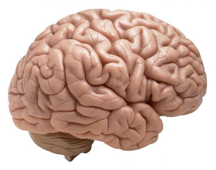
Summary: A new neuroimaging study reveals methylphenidate, better known as Ritalin, increases the level of dopamine available in the caudate. Along with increased dopamine levels, researchers also notice greater functional connectivity between the prefrontal cortex, hippocampus and precuneus, three areas of the brain associated with memory and attention.
Source: University of Wisconsin Madison.
There’s a reason so many children are prescribed methylphenidate, better known by the trade name Ritalin: it helps kids quell attention and hyperactivity problems and sit still enough to focus on a school lesson.
The drug keeps more of the neurotransmitter dopamine loose among brain cells, enhancing cell-to-cell transmission of information. We understand that, on a cellular level, according to Luis Populin. But we don’t know much more than that.
“It’s been said that we’re doing a large-scale, uncontrolled public health experiment on these kids,” says Populin, a University of Wisconsin-Madison neuroscience professor. “That’s a strong statement to make, but the reality is nobody can claim more than the cellular chemistry. Nobody can say: This is what are the consequences for the brains of kids that receive the drug.”
Populin and UW-Madison collaborators are beginning down that road, though, with the publication of a study this week in the Journal of Neuroscience describing increased connections between key parts of the brains of monkeys who have taken methylphenidate.
Populin and neuroscience researcher Abby Rajala gave three rhesus macaque monkeys a series of doses of methylphenidate — varying in size, but adjusted to mimic the doses typically prescribed to humans. They scanned the monkeys’ brains simultaneously with positron emission tomography (PET) able to indirectly track dopamine levels and functional magnetic resonance imaging (fMRI) that can reveal connections across the brain by showing areas working in conjunction.
Alexander Converse, a brain imaging specialist at UW-Madison’s Waisman Center, analyzed the PET data and found methylphenidate led to increases in levels of available dopamine in a central brain structure called the head of the caudate.
“It’s like political polling,” says Converse. “We give the monkeys a chemical tracer, which goes out and polls the receptors where the dopamine goes to see how many receptors are open. The less dopamine there is, the more receptors are available for the tracer.”
Looking at fMRI images taken simultaneously with the hike in dopamine tracked via PET, psychiatry Professor Rasmus Birn identified several areas of the brain experiencing increased connectivity with the head of the caudate.
“If we see two regions of the brain fluctuate in sync with each other, we call them functionally connected,” says Birn. “The assumption is that there’s a connection between the two. It doesn’t necessarily have to be direct, but the synced activity tells us there is communication between the two areas.”
With the methylphenidate-pegged increase in dopamine comes greater functional connectivity between the caudate and three brain structures called the prefrontal cortex, the hippocampus and the precuneus.
“These are areas that look relevant to the problems Ritalin is meant to address,” Birn says. “The prefrontal cortex is involved in sustained attention; the hippocampus plays a role in memory formation; the precuneus, in the back of the brain, is actually involved more in sensory motor function, and could be a way methylphenidate affects hyperactivity.”
Their findings are a first step toward understanding the way Ritalin affects the organization of the pathways that build brain networks used in attention and learning — and in what situations those alterations are helpful (or even whether they are harmful).
“It could be that the right dose to have your child sit quietly at their desk for an entire school day is actually having detrimental effects on other aspects of their cognitive function that are not as easily seen or measured,” Rajala says.

The next step is to see how varying amounts of the drug and changes in functional connectivity line up with performance on memory and learning tasks — to see, according to Rajala, whether differing doses expand or shut down different pathways to improve or hinder different brain functions.
“The ultimate goal is to empower physicians with better tools to decide if a child needs to receive a drug — and, if so, what type of drug and how much,” says Populin, whose work was supported by the university’s UW2020 initiative.
Key to the study was the addition of a simultaneous PET/MR scanner to the UW-Madison campus, and the ability to track dopamine and cross-brain connections at the same time. Methylphenidate’s effects shift person to person and even day to day, making the repeatability of the monkeys’ experiences and the scanning technology important.
“If you take a test with us today and come back to do it again tomorrow, your score may be very different because your physiological state may be different,” Populin says. “Maybe you had a crash with your car or you won a nice prize or you ate a bad breakfast. That’s enough to change your experience with methylphenidate, and what makes these methods an exciting way to show us what is happening in the brain.”
Source: Luis Populin, Rasmus Birn – University of Wisconsin Madison
Publisher: Organized by NeuroscienceNews.com.
Image Source: NeuroscienceNews.com image is in the public domain.
Original Research: Abstract for “Changes in endogenous dopamine induced by methylphenidate predict functional connectivity in non-human primates” by Rasmus M Birn, Alexander K Converse, Abigail Z Rajala, Andrew L Alexander, Walter F Block, Alan B McMillan, Bradley T Christian, Caitlynn N Filla, Dhanabalan Murali, Samuel A Hurley, Rick L Jenison and Luis C Populin in Journal of Neuroscience. Published December 10 2018.
doi:10.1523/JNEUROSCI.2513-18.2018
[cbtabs][cbtab title=”MLA”]University of Wisconsin Madison”Ritalin Drives Greater Connection Between Brain Areas Key to Memory and Attention.” NeuroscienceNews. NeuroscienceNews, 13 December 2018.
<https://neurosciencenews.com/ritalin-memory-attention-10335/>.[/cbtab][cbtab title=”APA”]University of Wisconsin Madison(2018, December 13). Ritalin Drives Greater Connection Between Brain Areas Key to Memory and Attention. NeuroscienceNews. Retrieved December 13, 2018 from https://neurosciencenews.com/ritalin-memory-attention-10335/[/cbtab][cbtab title=”Chicago”]University of Wisconsin Madison”Ritalin Drives Greater Connection Between Brain Areas Key to Memory and Attention.” https://neurosciencenews.com/ritalin-memory-attention-10335/ (accessed December 13, 2018).[/cbtab][/cbtabs]
Abstract
Changes in endogenous dopamine induced by methylphenidate predict functional connectivity in non-human primates
Dopamine (DA) levels in the striatum are increased by many therapeutic drugs, such as methylphenidate (MPH), which also alters behavioral and cognitive functions thought to be controlled by the prefrontal cortex (PFC) dose-dependently. We linked DA changes and functional connectivity (FC) using simultaneous [18F]fallypride PET and resting state functional magnetic resonance imaging (fMRI) in awake male rhesus monkeys after oral administration of various doses of MPH. We found a negative correlation between [18F]fallypride nondisplaceable binding potential (BPND) and MPH dose in the head of the caudate (hCd), demonstrating increased extracellular DA resulting from MPH administration. This decrease in BPND was negatively correlated with FC between hCd and PFC. Subsequent voxel-wise analyses revealed negative correlations with FC between the hCd and the dorsolateral PFC, hippocampus, and precuneus. These results, showing that MPH-induced changes in DA levels in hCd predict resting state FC, shed light on a mechanism by which changes in striatal DA could influence function in the PFC.
SIGNIFICANCE STATEMENT
Dopamine transmission is thought to play an essential role in shaping large scale-neural networks that underlie cognitive functions. It is the target of therapeutic drugs such as methylphenidate (Ritalin), which blocks the dopamine transporter, thereby increasing extracellular dopamine levels. Methylphenidate is used extensively to treat ADHD, even though its effects on cognitive functions and their underlying neural mechanisms are not well understood. To date, little is known about the link between changes in dopamine levels and changes in functional brain organization. Using simultaneous PET/MR imaging we show that methylphenidate-induced changes in endogenous dopamine levels in the head of the caudate predict changes in resting state functional connectivity between this structure and the prefrontal cortex, precuneus, and hippocampus.


