Summary: According to a new study, fear memory encoding is influences by parvalbumin interneurons in the amygdala.
Source: Mount Sinai Hospital.
Fear memory encoding, the process responsible for persistent reactions to trauma-associated cues, is influenced by a sparse but potent population of inhibitory cells called parvalbumin-interneurons (PV-INs) in the amygdala, according to a study conducted at the Icahn School of Medicine at Mount Sinai and published online July 14 in the journal Neuron.
The Mount Sinai study focused on identifying the synaptic connections between inhibitory PV-INs, sensory pathways and neighboring principal neurons in the basolateral amygdala, a brain region involved in detecting and responding to dangerous situations.
Stimuli encountered during a traumatic event can elicit strong emotional reactions long after the threat has subsided. These emotional memories are thought to be encoded through changes in the neural connections, or synapses, within the basolateral amygdala that provide outputs to other brain areas, controlling the so-called “fight or flight” response. These principal neurons increase their activity when an animal learns a threatening stimulus association. At other times, these cells are very quiet, despite their ongoing bombardment by sensory stimulation.
“Our study is the first to show that this default silencing may, in part, be attributable to a sparse population of inhibitory PV-INs,” says Roger Clem, PhD, Assistant Professor of Neuroscience and Psychiatry at the Icahn School of Medicine at Mount Sinai and lead investigator of the trial. “The complex anatomy of these cells may allow them to function like master regulators on a hair trigger, springing into action to suppress their neighbors when they detect even the slightest sensory perturbation.”
To investigate whether fear learning alters PV-IN properties and their silencing effect on surrounding neurons, the Mount Sinai team introduced fear conditioning in a mouse model, pairing an auditory tone with a subsequent aversive foot shock. They found that when animals acquire a fear memory, the suppressive influence of PV-INs is relieved, allowing the fear system to respond more vigorously when the auditory stimulus is re-encountered in order to trigger a fight-or-flight response.
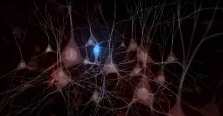
All sensations, movements, thoughts, memories and feelings are the result of signals that pass through nerve cells (neurons), the primary functional unit of the brain and central nervous system. When a signal passes from the cell body to the end of the cell axon that stretches away from the cell body, chemicals known as neurotransmitters are released into the synapse, the place where signals are exchanged between cells. The neurotransmitters then cross the synapse and attach to receptors on the neighboring cell, which can change the properties of the receiving cell.
Found throughout the brain and produced by neurons, gamma aminobutyric acid (GABA) is an inhibitory neurotransmitter that binds to GABA receptors, making the neighboring neuron less excitable. Fear-related disorders like anxiety and post-traumatic stress disorder (PTSD) are thought to partly result from an imbalance of excitatory and inhibitory nerve cells in the basolateral amygdala.
The current study team investigated GABA-synthesizing PV-INs after fear conditioning. Specifically, using electrophysiological and optogenetic techniques, the research team found that both input to and output from these cells was decreased after fear learning. In addition, the team found that memory encoding specifically affected PV-INs that respond most robustly to sensory input and that are thus uniquely positioned to regulate emotional reactivity.
The study findings indicate that inhibitory plasticity is a normal consequence of memory encoding and when dysregulated, it could lead to the hyper excitability of the amygdala, a hallmark of PTSD.
“Further investigation of the function of these PV-IN circuits under various stress paradigms may identify which of the processes we describe might be involved in pathological fear expression and ultimately, could establish cellular targets for reducing inappropriate amygdala responses and subsequent fear behaviors,” says Dr. Clem.
Funding: This research was supported by NIH grants MH105414 (R.L.C.) EY026053 (H.M.) and MH096678 (E.K.L.); a NARSAD Young Investigator Award (R.L.C.); and The Friedman Brain Institute at the Icahn School of Medicine at Mount Sinai.
Source: Elizabeth Dowling – Mount Sinai Hospital
Image Source: This NeuroscienceNews.com image is credited to Herry Lab.
Original Research: Abstract for “Multimodal and Site-Specific Plasticity of Amygdala Parvalbumin Interneurons after Fear Learning” by Elizabeth K. Lucas, Anita M. Jegarl, Hirofumi Morishita, and Roger L. Clem in Neuron. Published online July 14 2016 doi:10.1016/j.neuron.2016.06.032
[cbtabs][cbtab title=”MLA”]Mount Sinai Hospital. “Specialized Neurons in Brain Area Associated with Emotional Memory Play Role in Fear.” NeuroscienceNews. NeuroscienceNews, 14 July 2016.
<https://neurosciencenews.com/pv-ins-fear-4681/>.[/cbtab][cbtab title=”APA”]Mount Sinai Hospital. (2016, July 14). Specialized Neurons in Brain Area Associated with Emotional Memory Play Role in Fear. NeuroscienceNews. Retrieved July 14, 2016 from https://neurosciencenews.com/pv-ins-fear-4681/[/cbtab][cbtab title=”Chicago”]Mount Sinai Hospital. “Specialized Neurons in Brain Area Associated with Emotional Memory Play Role in Fear.” https://neurosciencenews.com/pv-ins-fear-4681/ (accessed July 14, 2016).[/cbtab][/cbtabs]
Abstract
Multimodal and Site-Specific Plasticity of Amygdala Parvalbumin Interneurons after Fear Learning
Highlights
•Lateral (LA) but not basal amygdala (BA) PV-INs receive potent afferent excitation
•PV-INs in LA but not BA mediate feedforward inhibition onto principal neurons
•Fear conditioning modulates synaptic input to PV-INs in a nucleus-specific manner
•PV-INs reduce GABA release onto LA principal neurons after fear conditioning
Summary
Stimulus processing in fear conditioning is constrained by parvalbumin interneurons (PV-INs) through inhibition of principal excitatory neurons. However, the contributions of PV-IN microcircuits to input gating and long-term plasticity in the fear system remain unknown. Here we interrogate synaptic connections between afferent pathways, PV-INs, and principal excitatory neurons in the basolateral amygdala. We find that subnuclei of this region are populated two functionally distinct PV-IN networks. PV-INs in the lateral (LA), but not the basal (BA), amygdala possess complex dendritic arborizations, receive potent excitatory drive, and mediate feedforward inhibition onto principal neurons. After fear conditioning, PV-INs exhibit nucleus- and target-selective plasticity, resulting in persistent reduction of their excitatory input and inhibitory output in LA but not BA. These data reveal previously overlooked specializations of amygdala PV-INs and indicate specific circuit mechanisms for inhibitory plasticity during the encoding of associative fear memories.
“Multimodal and Site-Specific Plasticity of Amygdala Parvalbumin Interneurons after Fear Learning” by Elizabeth K. Lucas, Anita M. Jegarl, Hirofumi Morishita, and Roger L. Clem in Neuron. Published online July 14 2016 doi:10.1016/j.neuron.2016.06.032