

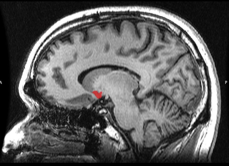
Summary: Researchers have discovered a connection between a lack of ovarian hormones and changes in the nucleus accembens following menopause.
Source: University of Missouri.
Possible treatments could improve motivation for physical activity.
As women enter menopause, their levels of physical activity decrease; for years scientists were unable to determine why. Now, researchers from the University of Missouri have found a connection between lack of ovarian hormones and changes in the brain’s pleasure center, a hotspot in the brain that processes and reinforces messages related to reward, pleasure, activity and motivation for physical exercise. Findings suggest that activation of brain receptors in that part of the brain may serve as a future treatment to improve motivation for physical activity in postmenopausal women.
“Postmenopausal women are more susceptible to weight gain and health issues,” said Victoria Vieira-Potter, assistant professor of nutrition and exercise physiology at MU. “This is especially frustrating for women, who already are dealing with significant changes to their bodies. We found that the decrease in physical activity that leads to weight gain may be caused by changes in brain activity.”
Vieira-Potter and her research team compared the physical activity of rats that were highly fit to rats that had lower fitness levels. The team studied the rats’ use of running wheels set up in the cages before and after the rats had their ovaries removed. They also examined gene expression changes of dopamine receptors within the brain’s pleasure center.
The high-fit rat group had more activity in the brain’s pleasure center, which correlated with greater wheel running before and after the loss of ovarian hormones. However, the high-fit rats still saw a significant reduction in wheel running after their ovaries were removed. This reduction in wheel running also correlated significantly with a reduction in their dopamine signaling levels, indicating that the brain’s pleasure center could be involved.
“We found that in both groups of rats, the hormonal changes from menopause led to changes in the brain that translated to less physical activity,” Vieira-Potter said. “The findings confirm previous evidence in humans and rodents that weight gain that occurs after menopause is likely due to decreased overall physical activity rather than increased energy intake from diet. Understanding what is causing the decrease in activity and subsequent weight gain may allow us to intervene, possibly by activating dopamine receptors, to preserve the motivation to be physically active.”
Jaume Padilla, assistant professor; and Jill Kanaley, professor and associate chair; in the department of nutrition and exercise physiology co-authored the study. Other contributors from MU were Young-Min Park, a former graduate student; Terese Zidon, graduate student; Rebecca Welly, lab manager in the department of nutrition and exercise physiology; Matthew Will, associate professor of psychological sciences in the College of Arts and Science and research investigator in the Bond Life Sciences Center; and Frank Booth, professor of biomedical sciences.
Researchers from the University of Michigan medical school and the University of Kansas medical center also contributed to the study.
Funding: “Effects of Intrinsic Aerobic Capacity and Ovariectomy on Voluntary Wheel Running and Nucleus Accumbens Dopamine Receptor Gene Expression,” recently was published in Physiology and Behavior. The research was supported by a MU Research Council Grant.
Source: Sandra Jacob – University of Missouri
Image Source: This NeuroscienceNews.com image is in the public domain
Original Research: Abstract for “Effects of Intrinsic Aerobic Capacity and Ovariectomy on Voluntary Wheel Running and Nucleus Accumbens Dopamine Receptor Gene Expression” by Young-Min Park, Jill A. Kanaley, Jaume Padilla, Terese Zidon, Rebecca J. Welly, Matthew J. Will, Steven L. Britton, Lauren G. Koch, Gregory N. Ruegsegger, Frank W. Booth, John P. Thyfault, and Victoria J. Vieira-Potter in Physiology and Behavior. Published online June 7 2016 doi:10.1016/j.physbeh.2016.06.006
[cbtabs][cbtab title=”MLA”]University of Missouri. “Deactivation of Brain Receptors in Postmenopausal Women May Lead to Lack of Physical Activity.” NeuroscienceNews. NeuroscienceNews, 1 August 2016.
<https://neurosciencenews.com/postmenopause-physical-activity-4765/>.[/cbtab][cbtab title=”APA”]University of Missouri. (2016, August 1). Deactivation of Brain Receptors in Postmenopausal Women May Lead to Lack of Physical Activity. NeuroscienceNew. Retrieved August 1, 2016 from https://neurosciencenews.com/postmenopause-physical-activity-4765/[/cbtab][cbtab title=”Chicago”]University of Missouri. “Deactivation of Brain Receptors in Postmenopausal Women May Lead to Lack of Physical Activity.” https://neurosciencenews.com/postmenopause-physical-activity-4765/ (accessed August 1, 2016).[/cbtab][/cbtabs]
Abstract
Effects of Intrinsic Aerobic Capacity and Ovariectomy on Voluntary Wheel Running and Nucleus Accumbens Dopamine Receptor Gene Expression
Rats selectively bred for high (HCR) and low (LCR) aerobic capacity show a stark divergence in wheel running behavior, which may be associated with the dopamine (DA) system in the brain. HCR possess greater motivation for voluntary running along with greater brain DA activity compared to LCR. We recently demonstrated that HCR are not immune to ovariectomy (OVX)-associated reductions in spontaneous cage (i.e. locomotor) activity. Whether HCR and LCR rats differ in their OVX-mediated voluntary wheel running response is unknown.
Purpose
To determine whether HCR are protected from OVX-associated reduction in voluntary wheel running.
Methods
Forty female HCR and LCR rats (age ~ 27 weeks) had either SHM or OVX operations, and given access to a running wheel for 11 weeks. Weekly wheel running distance was monitored throughout the intervention. Nucleus accumbens (NAc) was assessed for mRNA expression of DA receptors at sacrifice.
Results
Compared to LCR, HCR ran greater distance and had greater ratio of excitatory/inhibitory DA mRNA expression (both line main effects, P < 0.05). Wheel running distance was significantly, positively correlated with the ratio of excitatory/inhibitory DA mRNA expression across animals. In both lines, OVX reduced wheel running (P < 0.05). Unexpectedly, although HCR started with significantly greater voluntary wheel running, they had greater OVX-induced reduction in wheel running than LCR such that no differences were found 11 weeks after OVX between HCROVX and LCROVX (interaction, P < 0.05). This significant reduction in wheel running in HCR was associated with an OVX-mediated reduction in the ratio of excitatory/inhibitory DA mRNA expression.
Conclusion
The DA system in the NAc region may play a significant role in motivation to run in female rats. Compared to LCR, HCR rats run significantly more, which associates with greater ratio of excitatory/inhibitory DA mRNA expression. However, despite greater inherent motivation to run and an associated brain DA mRNA expression profile, HCR rats are not protected against OVX-induced reduction in wheel running or OVX-mediated reduction in the ratio of excitatory/inhibitory DA receptor mRNA expression. OVX-mediated reduction in motivated physical activity may be partially explained by a reduced ratio of excitatory/inhibitory DA receptor mRNA expression for which intrinsic fitness does not confer protection.
“Effects of Intrinsic Aerobic Capacity and Ovariectomy on Voluntary Wheel Running and Nucleus Accumbens Dopamine Receptor Gene Expression” by Young-Min Park, Jill A. Kanaley, Jaume Padilla, Terese Zidon, Rebecca J. Welly, Matthew J. Will, Steven L. Britton, Lauren G. Koch, Gregory N. Ruegsegger, Frank W. Booth, John P. Thyfault, and Victoria J. Vieira-Potter in Physiology and Behavior. Published online June 7 2016 doi:10.1016/j.physbeh.2016.06.006


