

Staying power soars at least 80-fold.
NIMH researchers have boosted the staying power of a social memory at least 80-fold by stimulating a circuit they discovered in mouse brain. A male mouse that would normally forget a female mouse it had just met within an hour instead remembered it at least a week later! Researchers precisely tweaked the circuit by genetically priming it to respond to pulses of light –a cutting edge technique called optogenetics. The study is the first to enhance social memory by stimulating a specific circuit.
The enhancement worked only if the male’s circuit had been stimulated while the memory was being formed, not recalled – and only during its first encounter with the female animal. The memory remained strong even after the male was distracted by the introduction of a second female mouse, which would normally degrade it.
“Someday, brain stimulation treatments might similarly target this pathway to help patients with declining social memories,” explained NIMH’s Scott Young, M.D., Ph.D. “The circuit allows the organism to remember and respond appropriately to social encounters, be they aversive or pleasant. If the same system is at work in humans, one can imagine how it would strengthen one’s memory for a bully to avoid in the future – or how it could be activated in ‘love at first sight’ to produce a lifelong memory.”
Young, Adam Smith, Ph.D., and colleagues in the NIMH intramural Section on Neural Gene Expression, Laboratory of Cellular and Molecular Regulation, report on their discovery January 5, 2016 in the journal Molecular Psychiatry.

Prior to the new study, Young’s team had shown that genetically silencing activity of a receptor for the social behavior-related hormone vasopressin blocks social memory. They also knew that brain expression of this vasopressin 1b receptor is confined mostly to a little studied part of the memory hub, or hippocampus, called CA2 – and that blocking CA2 reduces social memory. So the researchers set out to discover the upstream circuitry that triggers release of vasopressin in CA2.
A prime candidate was a set of neurons that project to CA2 from a part of the hypothalamus called the PVN (paraventricular nucleus). The PVN integrates information from external and internal environments to orchestrate stress responses.
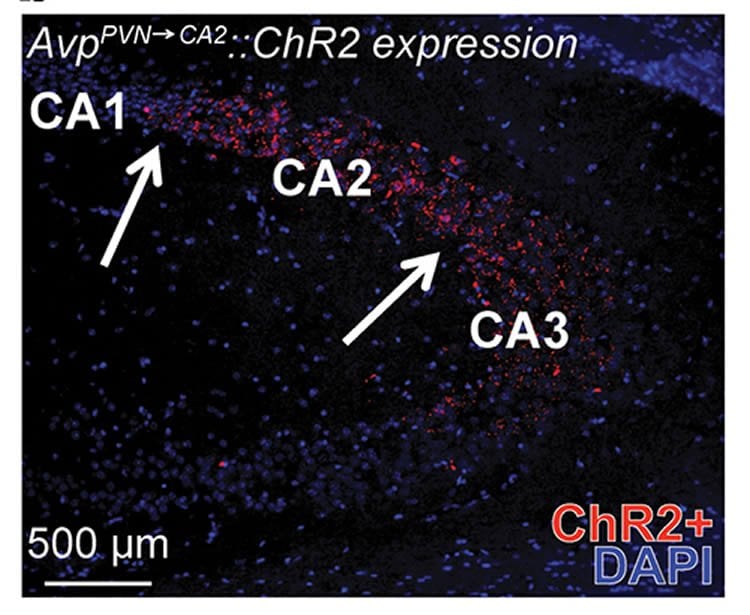
As suspected, pathway tracing revealed a single, anatomically distinct circuit from the PVN that releases vasopressin in CA2. The researchers used implanted LED fiber optics to stimulate the circuit – genetically engineered to be light-activated – while male mice sniffed out novel females. When exposed to the same female later, less sniffing signaled that the male remembered it. As noted, such memories persisted at least 80 times longer-than-normal in optogenetically-stimulated males and resisted weakening effects of distractions. Chemically blocking vasopressin activity in CA2 blocked the stimulated social memory enhancement.
This confirmed that vasopressin activity in CA2, triggered by the circuit from PVN, is a key player in social memory – although other vasopressin pathways are also likely involved.
“If circuitry similar to what we’re seeing in mice turns out to be also at work in the human brain, treatments based on similar targeted brain pathway stimulation might someday help to improve the relationships of people experiencing social memory impairment due to dementias and mental illnesses,” said Young.
Source: Jules Asher – NIMH/NIH
Image Credit: The images re credited to Adam Smith Ph.D., NIMH
Original Research: Abstract for “Targeted activation of the hippocampal CA2 area strongly enhances social memory” by A S Smith, S K Williams Avram, A Cymerblit-Sabba, J Song and W S Young in PNAS. Published online January 5 2016 doi:10.1038/mp.2015.189
Abstract
Targeted activation of the hippocampal CA2 area strongly enhances social memory
Social cognition enables individuals to understand others’ intentions. Social memory is a necessary component of this process, for without it, subsequent encounters are devoid of any historical information. The CA2 area of the hippocampus, particularly the vasopressin 1b receptor (Avpr1b) expressed there, is necessary for memory formation. We used optogenetics to excite vasopressin terminals, originating from the hypothalamic paraventricular nucleus, in the CA2 of mice. This markedly enhanced their social memory if the stimulation occurred during memory acquisition, but not retrieval. This effect was blocked by an Avpr1b antagonist. Finally, this enhanced memory is resistant to the social distraction of an introduced second mouse, important for socially navigating populations of individuals. Our results indicate the CA2 can increase the salience of social signals. Targeted pharmacotherapy with Avpr1b agonists or deep brain stimulation of the CA2 are potential avenues of treatment for those with declining social memory as in various dementias.
“Targeted activation of the hippocampal CA2 area strongly enhances social memory” by A S Smith, S K Williams Avram, A Cymerblit-Sabba, J Song and W S Young in PNAS. Published online January 5 2016 doi:10.1038/mp.2015.189


