

Water-based imaging technique maps brain neurons prior to surgery.
Neurosurgeons at UC San Diego Health System are using a new approach to visualize the brain’s delicate anatomy prior to surgery. The novel technique allows neurosurgeons to see the brain’s nerve connections thus preserving and protecting critical functions such as vision, speech and memory. No needles, dyes or chemicals are needed to create the radiology scan. The main imaging ingredient? Water.
“The brain can be mapped by tracking the movement of its water molecules,” said Clark Chen, MD, PhD, neurosurgeon and vice-chairman of neurosurgery at UC San Diego Health System. “Water molecules in brain nerves move in an oriented manner. However, outside the nerves, the molecules move randomly. Neurosurgeons at UC San Diego can use these distinct properties to locate important connections and to guide where surgery should occur or not.”
The technique, called tractography or diffusion tensor imaging (DTI), has been used for investigational and diagnostic purposes to better understand the effect of stroke and neurological diseases, such as Alzheimer’s. UC San Diego Health System neurosurgeons are among the first in the nation to apply this technology to guide brain tumor surgery.
“There are no margins for error in the brain. Every centimeter of brain tissue contains millions of neural connections so every millimeter counts,” said Chen. “With tractography, we can visualize the most important of these connections to avoid injury. In doing so, we will preserve the quality of life for our patients with brain cancer.”
Anthony Chetti is one of the beneficiaries of tractography-guided brain surgery. Chetti developed a tumor in the region of the brain called the occipital lobe, the portion of the brain responsible for processing visual information.
“Anytime that you are told that you can potentially lose your vision, you are scared,” said Chetti, a San Diego school teacher. “But when Dr. Chen shared the tractography images with me and showed me how he was going to avoid injury to the connection between my eye and the occipital lobe, I was reassured.”
Chetti underwent a complete excision of the brain tumor without any damage to his vision.
“When I woke up from surgery, I asked for my glasses immediately and began running systems checks. I could see the clock. I could read the words on a sign. It was immediately evident that there were no problems,” said Chetti.
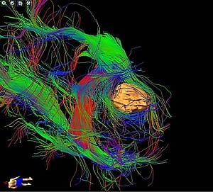
Tractography scans can reveal tiny open paths between nerve fibers to reach brain tumors. The scans are color coded and display tiny neural connections. Other current imaging techniques such as computed tomography (CT) and conventional magnetic resonance imaging (MRI) cannot achieve this type of visual display.
“There is no way of predicting where these fibers would lie except with tractography,” said Chen. “I believe that many brain tumor patients will benefit from tractography-guided surgery. By observing the flow of water molecules, we can visually reconstruct the complex symphony of brain connections. The resulting images are not only accurate but breathtakingly beautiful, giving us a glimpse into the extraordinary human mystery of the brain.”
Chen added that this radiology application is new and that UC San Diego teams are learning how best to apply the technology. Chen is actively investigating ways to optimize the use of tractography information through collaboration with world renowned experts, including Anders M. Dale, PhD, vice-chair of radiology, and director of the newly established Center for Translational Imaging and Personalized Medicine at UC San Diego, School of Medicine.
Notes about this neuroimaging and brain mapping research
Contact: Jackie Carr – UCSD Health System
Source: UCSD Health System press release
Image Source: The image is adapted from the UCSD Health System press release.
Video Source: The video “Advances in Imaging Brain Tumors at UC San Diego” is available at the UCSD Health System YouTube page.
#neuroimaging, #neurosciencevideos, #neurology, #brainmapping


