

Summary: Renewed, thin myelin sheaths are sufficient to help restore nervous system impairments in diseases like multiple sclerosis, a new study in PNAS reports.
Source: University of Wisconsin Madison.
In the central nervous system of humans and all other mammals, a vital insulating sheath composed of lipids and proteins around nerve fibers helps speed the electrical signals or nerve impulses that direct our bodies to walk, talk, breathe, swallow or perform any routine physical act.
But diseases of the nervous system, including multiple sclerosis (MS) in people, degrade this essential insulation known as myelin, disrupting the flow of information between the brain and the body, impairing movement, dimming vision and blunting the ability to function normally.
And while scientists have long studied myelin and understand its role in disease when it degrades, they have puzzled over how myelin repairs itself naturally and whether the thinned sheaths that are a hallmark of the healing nervous system are adequate for restoring the brain’s circuitry over the long haul.
This week (Oct. 23, 2017), in a study published in the Proceedings of the National Academy of Sciences, a team of researchers from the University of Wisconsin-Madison reports that in long-lived animals, renewed but thin myelin sheaths are enough to restore the impaired nervous system and can do so for years after the onset of disease.
The team’s findings reinforce the idea that thin myelin sheaths are a valid, persistent marker of remyelination, a hypothesis challenged by other recent research. “As the only biomarker of myelin repair available this would leave us without any means of identifying or quantifying myelin repair,” explains Ian Duncan, an expert on demyelinating diseases at the UW-Madison School of Veterinary Medicine and the senior author of the new study.
Duncan and his team looked at a unique genetic disorder that naturally afflicts Weimaraners, a breed of dog that as 12- to 14-day-old pups develop a severe tremor and loss of coordination. The condition is known to occur as the development of the myelin sheath in parts of the dog’s central nervous system is delayed. The symptoms gradually diminish and in most cases disappear altogether by 3-4 months of age.
“This is a very widespread mutation in the breed,” says Duncan, noting that myelin repair mimicking what is seen in remyelination is known to occur in these dogs as the rejuvenated nerve fibers have a thinned myelin sheath.
The new Wisconsin study was made possible as 13 years ago two Weimaraner pups, littermates, were seen as patients at the School of Veterinary Medicine and Duncan was able to maintain contact with the owners after the dogs were adopted and retrieve samples of spinal tissue after the dogs lived out their lives. As they aged, the dogs exhibited few signs of tremor and were deemed ‘neurologically normal’ up to 13 years of age.
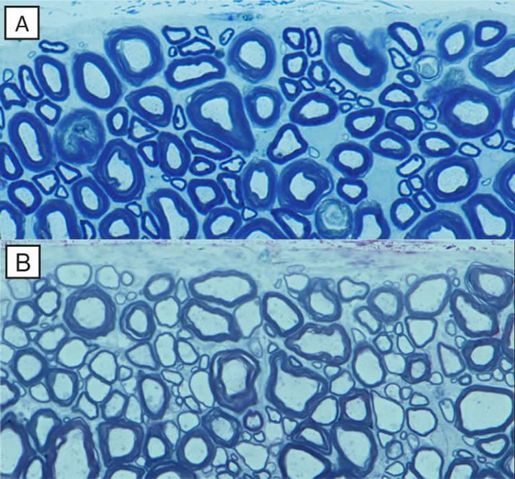
The purpose of the study, says Duncan, was to confirm that thin myelin sheaths persisted and supported normal neurologic function.
To expand on the results, Duncan also looked at a condition in cats, another long-lived species that has been shown to fully recover nervous system function after demyelination. In particular, Duncan’s team was interested in remyelination of the optic nerves.
That element of the study, looking at remyelination two years after the onset of the condition, Duncan notes, is an example of “true demyelination and remyelination. We found that nearly every optic nerve fiber was remyelinated with a thin myelin sheath, which is important for understanding human disease because in multiple sclerosis, the optic nerve is often the first to be demyelinated.”
The new findings confirm that the gold standard for evaluating remyelination is the long-term persistence of thin myelin sheaths, which support nerve fiber function and survival, Duncan notes. The results are important for diseases like MS as it means that new therapies designed to promote myelin repair can be safely evaluated and quantified based on the presence of thin myelin sheaths.
Funding: These studies were supported in part by NMSS grant RG-1501-02876 and by a prior grant from the MS Hope for a Cure Foundation.
Source: Ian Duncan – University of Wisconsin Madison
Publisher: Organized by NeuroscienceNews.com.
Image Source: NeuroscienceNews.com image is credited to the researchers.
Original Research: Abstract for “Thin myelin sheaths as the hallmark of remyelination persist over time and preserve axon function” by Ian D. Duncan, Rachel L. Marik, Aimee T. Broman, and Moones Heidari in PNAS. Published online October 24 2017 doi:10.1073/pnas.1714183114
[cbtabs][cbtab title=”MLA”]University of Wisconsin Madison “A Little Myelin Goes A Long Way To Restore Nervous System Function.” NeuroscienceNews. NeuroscienceNews, 24 October 2017.
<https://neurosciencenews.com/myelin-nervous-system-7802/>.[/cbtab][cbtab title=”APA”]University of Wisconsin Madison (2017, October 24). A Little Myelin Goes A Long Way To Restore Nervous System Function. NeuroscienceNews. Retrieved October 24, 2017 from https://neurosciencenews.com/myelin-nervous-system-7802/[/cbtab][cbtab title=”Chicago”]University of Wisconsin Madison “A Little Myelin Goes A Long Way To Restore Nervous System Function.” https://neurosciencenews.com/myelin-nervous-system-7802/ (accessed October 24, 2017).[/cbtab][/cbtabs]
Abstract
Thin myelin sheaths as the hallmark of remyelination persist over time and preserve axon function
The presence of thin myelin sheaths in the adult CNS is recognized as a marker of remyelination, although the reason there is not a recovery from demyelination to normal myelin sheath thickness remains unknown. Remyelination is the default pathway after myelin loss in all mammalian species, in both naturally occurring and experimental disease. However, there remains uncertainty about whether these thin sheaths thicken with time and whether they remain viable for extended periods. We provide two lines of evidence here that thin myelin sheaths may persist indefinitely in long-lived animal models. In the first, we have followed thin myelin sheaths in a model of delayed myelination during a period of 13 years that we propose results in the same myelin sheath deficiencies as seen in remyelination; that is, thin myelin sheaths and short internodes. We show that the myelin sheaths remain thin and stable on many axons throughout this period with no detrimental effects on axons. In a second model system, in which there is widespread demyelination of the spinal cord and optic nerves, we also show that thinly remyelinated axons with short internodes persist for over the course of 2 y. These studies confirm the persistence and longevity of thin myelin sheaths and the importance of remyelination to the long-term health and function of the CNS.
“Thin myelin sheaths as the hallmark of remyelination persist over time and preserve axon function” by Ian D. Duncan, Rachel L. Marik, Aimee T. Broman, and Moones Heidari in PNAS. Published online October 24 2017 doi:10.1073/pnas.1714183114


