

Summary: Melanocyte stem cells from human hair follicles that carry CD34 have the ability to turn into glial cells. The CD34+ stem cells can regenerate myelin, both on neurons and in mouse models with a genetic defect that prevents the formation of healthy myelin sheaths. The findings could have positive implications for the treatment of demyelinating diseases, such as Multiple Sclerosis.
Source: PLOS
A subset of the stem cells in hair follicles have the potential to regenerate the coating that insulates neurons in mice, reports Thomas Hornyak of the VA Maryland Health Care System and the University of Maryland School of Medicine and colleagues, in a new study published 24th April in PLOS Genetics. The study offers a new direction for finding therapeutic options for certain neurodegenerative diseases.
Hair and skin take on varying shades of red, brown, black and yellow due to the pigments produced by cells called melanocytes. Melanocytes originate embryonically from cells called neural crest cells, which are cells that can also give rise to neurons and their supporting glial cells. Previously, Hornyak and colleagues identified two different pockets of stem cells that create melanocytes inside mature hair follicles. In the current study, they show that the two groups of the melanocyte stem cells can be identified and separated based on whether they are coated in a glycoprotein called CD34, a surface molecule which is present on other types of stem cells, including stem cells of the blood.
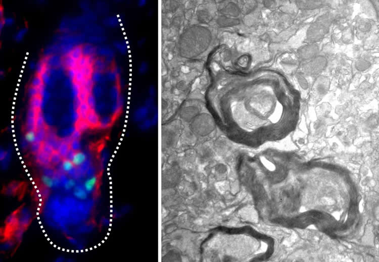
Using hair follicles from mice, the researchers isolated the two populations of melanocyte stem cells and grew them in culture. They were surprised to find that the cells carrying CD34 turn into glial cells. In the nervous system, glial cells coat neurons with a fatty insulation called myelin, which increases the speed that nerve impulses can travel. Furthermore, the researchers discovered that the CD34-positive stem cells could regenerate myelin on neurons, both in cell cultures and when injected into mice carrying a genetic defect that prevents them from forming myelin sheaths.
The new findings suggest that the pocket of CD34-positive melanocyte stem cells in the hair follicle retain some of their earlier abilities. If similar populations exist in human hair follicles, they potentially could be tapped to develop new treatments for nerve injuries and for demyelinating diseases, such as multiple sclerosis. “In the future, we plan to continue our research in this area by determining whether these cells can enhance functional recovery from neuronal injury,” said author Dr. Thomas Hornyak, “and leverage genome-wide information we have described in the current study to identify similar cells in human skin.
Funding: This research was supported in part by NIH/NIAMS R01 grant 1R01AR064810, United States Department of Health & Human Services; VA Merit Award BX002582, Biomedical Laboratory Research & Development, Office of Research & Development, United States Department of Veterans Affairs; Dean’s Office funds, University of Maryland School of Medicine; and the Baltimore Research and Education Foundation, VA Maryland Health Care System. The funders had no role in study design, data collection and analysis, decision to publish, or preparation of the manuscript.
Competing Interests: The authors declare no competing financial interests.
Source:
PLOS
Media Contacts:
Thomas J Hornyak – PLOS
Image Source:
The image is credited to Sandeep Joshi, University of Maryland School of Medicine.
Original Research: Open access.
“CD34 defines melanocyte stem cell subpopulations with distinct regenerative properties”
Sandeep S. Joshi, Bishal Tandukar, Li Pan, Jennifer M. Huang, Ferenc Livak, Barbara J. Smith, Theresa Hodges, Anup A. Mahurkar, Thomas J. Hornyak. PLOS Genetics. doi:10.1371/journal.pgen.1008034
Abstract
CD34 defines melanocyte stem cell subpopulations with distinct regenerative properties
Melanocyte stem cells (McSCs) are the undifferentiated melanocytic cells of the mammalian hair follicle (HF) responsible for recurrent generation of a large number of differentiated melanocytes during each HF cycle. HF McSCs reside in both the CD34+ bulge/lower permanent portion (LPP) and the CD34- secondary hair germ (SHG) regions of the HF during telogen. Using Dct-H2BGFP mice, we separate bulge/LPP and SHG McSCs using FACS with GFP and anti-CD34 to show that these two subsets of McSCs are functionally distinct. Genome-wide expression profiling results support the distinct nature of these populations, with CD34- McSCs exhibiting higher expression of melanocyte differentiation genes and with CD34+ McSCs demonstrating a profile more consistent with a neural crest stem cell. In culture and in vivo, CD34- McSCs regenerate pigmentation more efficiently whereas CD34+ McSCs selectively exhibit the ability to myelinate neurons. CD34+ McSCs, and their counterparts in human skin, may be useful for myelinating neurons in vivo, leading to new therapeutic opportunities for demyelinating diseases and traumatic nerve injury.


