

Summary: Combining advanced microscopy and mathematical modeling, researchers have discovered a pattern that governs the growth of neurons.
Source: Stanford
Life is rife with patterns. It’s common for living things to create a repeating series of similar features as they grow: think of feathers that vary slightly in length on a bird’s wing or shorter and longer petals on a rose..
It turns out the brain is no different. By employing advanced microscopy and mathematical modeling, Stanford researchers have discovered a pattern that governs the growth of brain cells or neurons. Similar rules could guide the development of other cells within the body, and understanding them could be important for successfully bioengineering artificial tissues and organs.
Their study, published in Nature Physics, builds on the fact that the brain contains many different types of neurons and that it takes several types working in concert to perform any tasks. The researchers wanted to uncover the invisible growth patterns that enable the right kinds of neurons to arrange themselves into the right positions to build a brain.
“How do cells with complementary functions arrange themselves to construct a functioning tissue?” said study co-author Bo Wang, an assistant professor of Bioengineering. “We chose to answer that question by studying a brain because it had been commonly assumed that the brain was too complex to have a simple patterning rule. We surprised ourselves when we discovered there was, in fact, such a rule.”
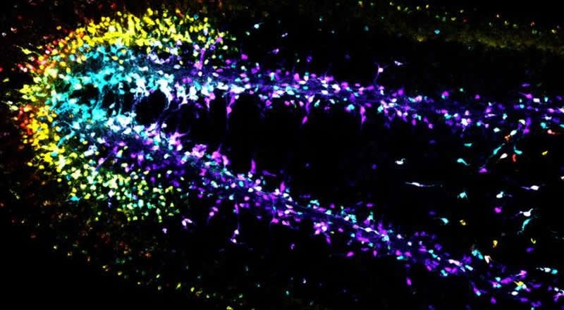
The brain they chose to examine belonged to a planarian, a millimeter-long flatworm that can regrow a new head every time after amputation. First, Wang and Margarita Khariton, a graduate student in his lab, used fluorescent stains to mark different types of neurons in the flatworm. They then used high-resolution microscopes to capture images of the whole brain – glowing neurons and all – and analyzed the patterns to see if they could extract from them the mathematical rules guiding their construction.
What they found was that each neuron is surrounded by roughly a dozen neighbors similar to itself, but that interspersed among them are other kinds of neurons. This unique arrangement means that no single neuron sits flush against its twin, while still allowing different types of complementary neurons to be close enough to work together to complete tasks.
The researchers found that this pattern repeats over and over across the entire flatworm brain to form a continuous neural network. Study co-authors Jian Qin, an assistant professor of chemical engineering, and postdoctoral scholar Xian Kong developed a computational model to show that this complex network of functional neighborhoods stems from the tendency of neurons to pack together as closely as possible without being too close to other neurons of the same type.
While neuroscientists might someday adapt this methodology to study neuronal patterning in the human brain, the Stanford researchers believe the technique could be more usefully applied to the emerging field of tissue engineering.
The basic idea is simple: tissue engineers hope to induce stem cells, the powerful, general-purpose cells from which all cell types derive, to grow into the various specialized cells that form a liver, kidney or heart. But scientists will need to arrange those diverse cells into the right patterns if they want the heart to beat.
“The question of how organisms grow into forms that carry out useful functions has fascinated scientists for centuries,” Wang said. “In our technological era, we are not limited to understanding these growth patterns at the cellular level but can also find ways to implement these rules for bioengineering applications.”
Source:
Stanford
Media Contacts:
Tom Abate – Stanford
Image Source:
The image is credited to Wang Lab.
Original Research: Closed access
“Chromatic neuronal jamming in a primitive brain”. Margarita Khariton, Xian Kong, Jian Qin & Bo Wang.
Nature Physics doi:10.1038/s41567-020-0809-9.
Abstract
Chromatic neuronal jamming in a primitive brain
Jamming models developed in inanimate matter have been widely used to describe cell packing in tissues but predominantly neglect cell diversity, despite its prevalence in biology. Most tissues, animal brains in particular, comprise a mix of many cell types, with mounting evidence suggesting that neurons can recognize their respective types as they organize in space. How cell diversity revises the current jamming paradigm is unknown. Here, in the brain of the flatworm planarian Schmidtea mediterranea, which exhibits remarkable tissue plasticity within a simple, quantifiable nervous system we identify a distinct packing state, ‘chromatic’ jamming. Combining experiments with computational modelling, we show that neurons of distinct types form independent percolating networks barring any physical contact. This jammed state emerges as cell packing configurations become constrained by cell type-specific interactions and therefore may extend to describe cell packing in similarly complex tissues composed of multiple cell types.


