

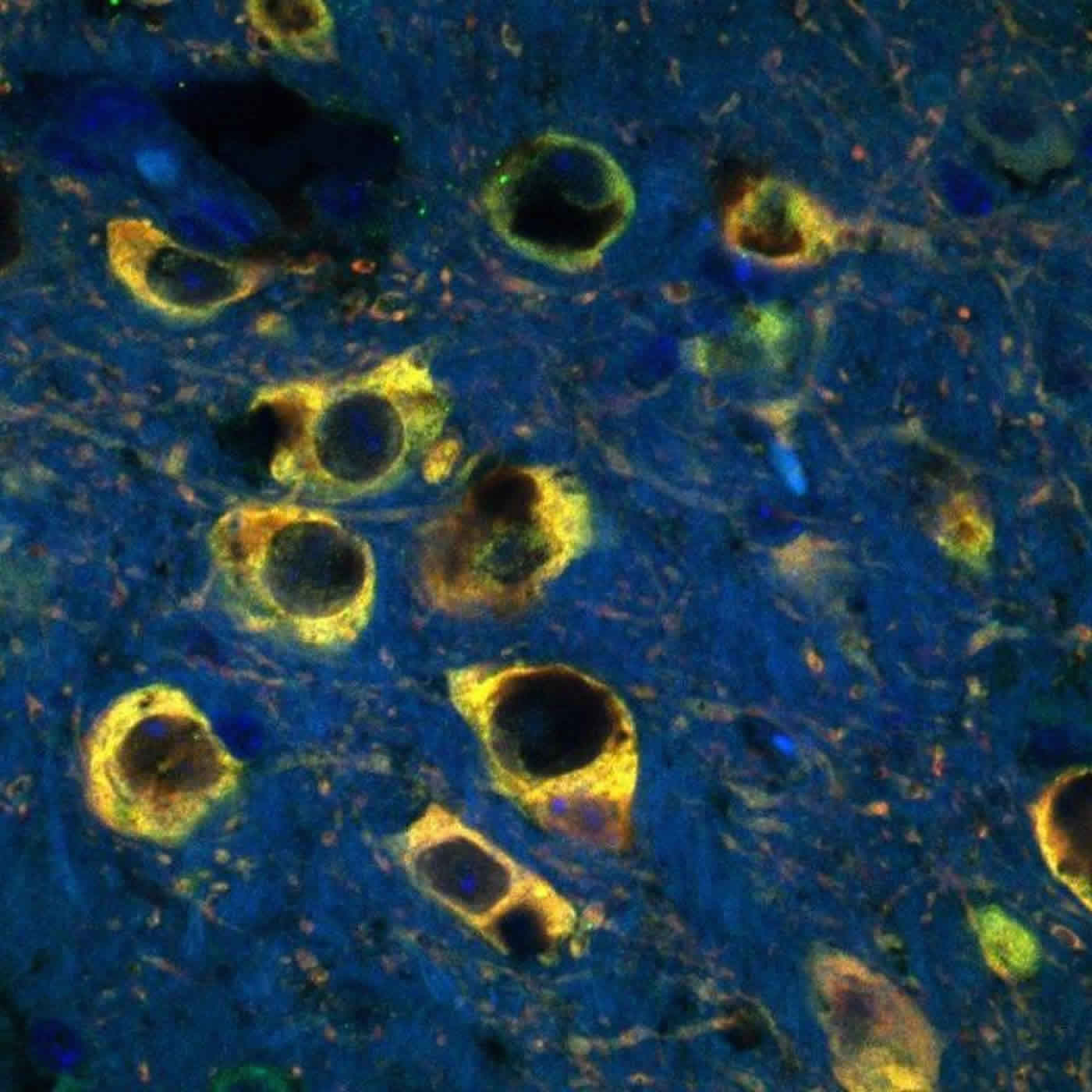
Excess iron impairs cellular recycling resulting in toxic oxidative stress.
It’s long been known that excess iron is found in the brains of patients with Parkinson’s disease (PD), an incurable neurodegenerative condition that affects motor function. The mechanism by which the iron wreaks damage on neurons involved in PD has not been clear. Research from the Andersen lab at the Buck Institute suggests that the damage stems from an impairment in the lysosome, the organelle that acts as a cellular recycling center for damaged proteins. Scientists report the impairment allows excess iron to escape into the neurons where it causes toxic oxidative stress. The research will be published online in The Journal of Neuroscience on Jan. 27, 2016.
Lysosomes are key to a process called autophagy, whereby damaged proteins are broken down into building blocks that are used to make newly-built proteins to take their place. It’s the cellular equivalent of recycling. With age, the ability of the lysosome to participate in autophagy becomes slower, resulting in the build-up of non-protein “garbage” within the cells. Less-than-optimal autophagy has been associated with several age-related diseases, including PD.
“It’s recently been realized that one of the most important functions of the lysosome is to store iron in a place in the cell where it is not accessible to participate in toxic oxidative stress-producing reactions,” said Julie K. Andersen, PhD, senior scientist and Buck Institute faculty. “Now we have demonstrated that a mutation in a lysosomal gene results in the toxic release of iron into the cell resulting in neuronal cell death.”
Spearheaded by staff scientist Shankar J. Chinta, PhD, the work (done in both mice and cultured human dopaminergic cells) involved a mutation in a gene (ATP13A2) associated with a rare early onset form of PD called Kufor-Rakeb syndrome. When researchers knocked out ATP13A2 the lysosome was unable to maintain the balance of iron within the cell.
The mutation responsible for Kufor-Rakeb was identified in 2010. Those suffering from the condition, which is named for the village in Jordan where the syndrome was first described, experience disease onset in adolescence. “Mutations in this same gene have also been recently linked to sporadic forms of PD,” said Andersen. “This suggests that age-related impairments in lysosomal function that impact the ability of neurons to maintain a healthy balance of iron are part of what underlies the presentation of PD in the general population.”
Andersen has a long-standing interest in the role of excess iron in PD and this current work provides an example of the value of basic research in drug discovery. In 2003 her lab showed that tying up excess iron with a metal chelator (derived from the Greek word for claw) protected mice from the ravaging effects of the well-known Parkinson’s inducing toxin, MPTP. The study provided an important link between the observed excessive iron in the brains of PD patients and oxidative stress associated with neurodegeneration. “The issue with iron chelation is that it’s a sledge hammer — it pulls iron from the cells indiscriminately and iron is needed throughout the body for many biological functions,” said Andersen. “Now we have a more specific target that we can hit with a smaller hammer, which could allow us to selectively impact iron toxicity within the affected neurons.”
Other Buck scientists involved in the study include Subramanian Rajagopalan and Anand Rane. This work was supported by the National Institutes of Health (RO1 NS047198, NS047198, NS041264, and AG012141).
Source: Kris Rebillot – Buck Institute for Research on Aging
Image Source: The image is credited to Subramanian Rajagopalan, MSc. Buck Institute for Research on Aging
Original Research: Abstract for “Regulation of ATP13A2 via PHD2-HIF1α Signaling Is Critical for Cellular Iron Homeostasis: Implications for Parkinson’s Disease” by Subramanian Rajagopalan, Anand Rane, Shankar J. Chinta, and Julie K. Andersen in Journal of Neuroscience. Published online January 27 2016 doi:10.1523/JNEUROSCI.3117-15.2016
Abstract
Regulation of ATP13A2 via PHD2-HIF1α Signaling Is Critical for Cellular Iron Homeostasis: Implications for Parkinson’s Disease
We previously reported that pharmacological inhibition of a class of enzymes known as prolyl hydroxylase domain proteins (PHDs) has neuroprotective effects in various in vitro and in vivo models of Parkinson’s disease (PD). We hypothesized that this was due to inhibition of the PHD2 isoform, preventing it from hydroxylating the transcription factor hypoxia inducible factor 1 α (HIF1α), targeting it for eventual proteasomal degradation. HIF1α itself induces the transcription of various cellular stress genes, including several involved in iron metabolism. Although all three isoforms of PHD are expressed within vulnerable dopaminergic (DAergic) substantia nigra pars compacta neurons, only select downregulation of the PHD2 isoform was found to protect against in vivo neurodegenerative effects associated with the mitochondrial neurotoxin 1-methyl-4-phenyl-1,2,3,6-tetrahydropyridine. These findings were corroborated in induced pluripotent stem cell-derived neurons, providing validation in a pertinent human cell model. PHD2 inhibition was found to result in increased expression of ATP13A2, mutation of which is responsible for a rare juvenile form of PD known as Kufor-Rakeb syndrome. Knockdown of ATP13A2 expression within human DAergic cells was found to abrogate restoration of cellular iron homeostasis and neuronal cell viability elicited by inhibition of PHD2 under conditions of mitochondrial stress, likely via effects on lysosomal iron storage. These data suggest that regulation of ATP13A2 by the PHD2-HIF1α signaling pathway affects cellular iron homeostasis and DAergic neuronal survival. This constitutes a heretofore unrecognized process associated with loss of ATP13A2 function that could have wide-ranging implications for it as a therapeutic target for PD and other related conditions.
SIGNIFICANCE STATEMENT Reductions in PHD2 activity within dopaminergic neurons in vivo and in cultured human induced pluripotent stem cell-derived neurons protects against mitochondrial stress-induced neurotoxicity. Protective effects are dependent on downstream HIF-1α expression. Knockdown of ATP13A2, a gene linked to a rare juvenile form of Parkinson’s disease and recently identified as a novel HIF1α target, was found to abrogate maintenance of cellular iron homeostasis and neuronal viability elicited by PHD2 inhibition in vivo and in cultured dopaminergic cells under conditions of mitochondrial stress. Mechanistically, this was due to ATP13A2’s role in maintaining lysosomal iron stores. This constitutes a novel mechanism by which alterations in ATP13A2 activity may be driving PD-related neuropathology.
“Regulation of ATP13A2 via PHD2-HIF1α Signaling Is Critical for Cellular Iron Homeostasis: Implications for Parkinson’s Disease” by Subramanian Rajagopalan, Anand Rane, Shankar J. Chinta, and Julie K. Andersen in Journal of Neuroscience. Published online January 27 2016 doi:10.1523/JNEUROSCI.3117-15.2016


