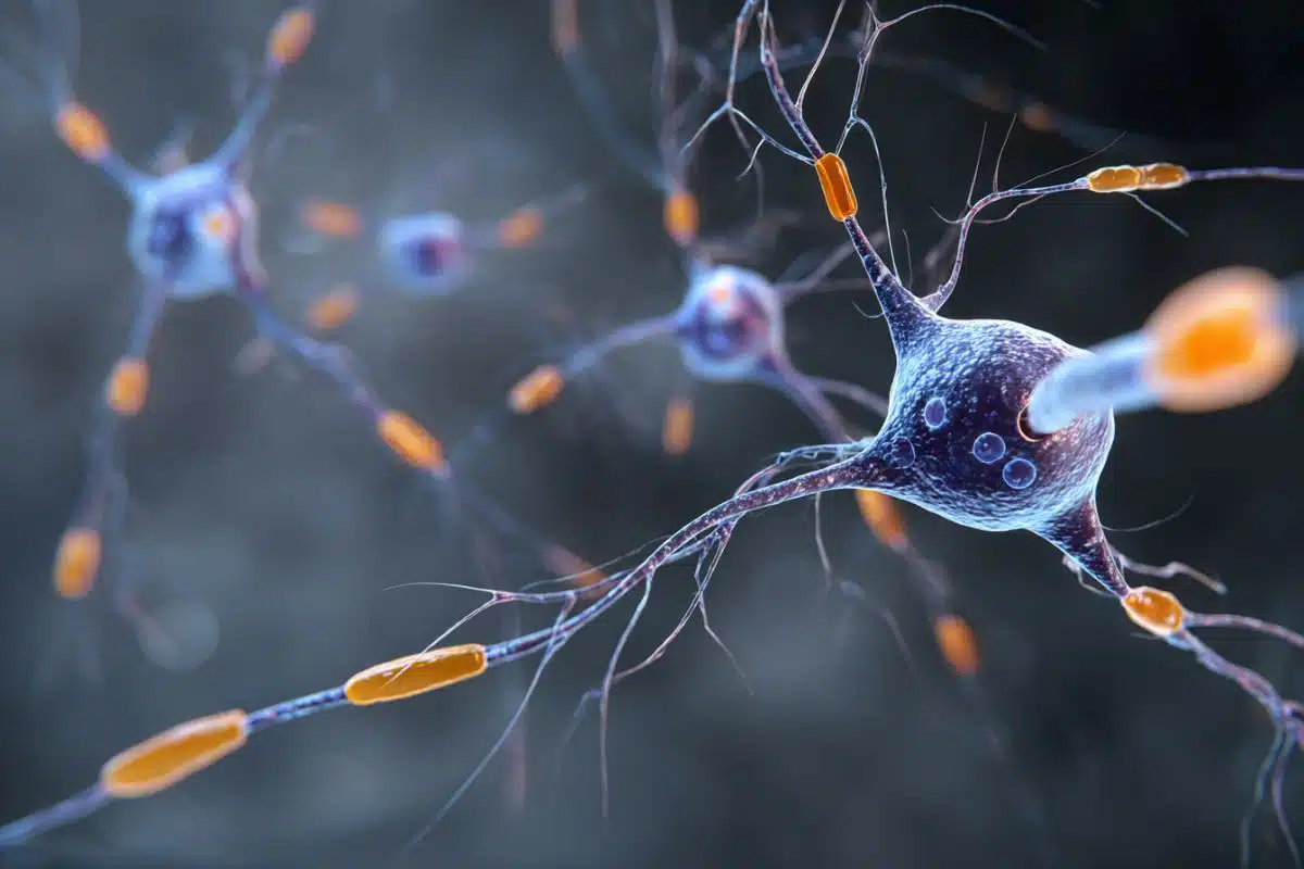
Summary: A new study uncovered a structural explanation for why women are disproportionately vulnerable to Alzheimer’s disease (AD). The research shifts focus away from traditional brain cells to investigate the extracellular matrix (ECM), the foundational, non-cellular “mortar” filling the spaces between brain cells.
By analyzing mouse models with localized estrogen depletion, investigators discovered that the combination of aging, female sex, and post-menopausal estrogen loss triggers an architectural collapse within the hippocampal ECM, dismantling the supportive scaffold required for memory retention.
Key Facts
- The Female Vulnerability Gap: Nearly two-thirds of all Americans living with Alzheimer’s disease are women. While scientists have long hypothesized that post-menopausal estrogen decline robs the female brain of its natural neuroprotective shield, the precise biological mechanisms have remained poorly understood.
- The Intercellular Scaffold: Making up nearly 20% of total brain volume, the extracellular matrix (ECM) functions like mortar between bricks, creating a highly abundant molecular network and supportive scaffold in the hippocampus that allows brain cells to communicate and function.
- The Localized Estrogen Shift: Prior to menopause, a woman’s ovaries serve as the primary engine for estrogen production. Post-menopause, systemic levels plummet sharply, leaving the brain as a primary site for local estrogen synthesis—a localized supply that is severely diminished in female Alzheimer’s patients.
- Dismantling the Space Between Cells: By deploying genetically engineered mouse models lacking aromatase (the essential enzyme required to synthesize estrogen), the Northwestern team isolated the effects of aging and estrogen loss across both sexes. They discovered that older females are uniquely sensitive to this brain-estrogen crash, which causes widespread degradation of the ECM.
- Challenging Anti-Amyloid Interventions: Modern frontline Alzheimer’s therapeutics, such as lecanemab and donanemab, focus strictly on clearing abnormal amyloid protein buildups. However, their real-world ability to slow memory loss or restore daily cognitive function remains highly controversial and mixed.
- A New Therapeutic Horizon: Senior authors Dr. Hong Zhao and Dr. Serdar Bulun note that these findings present a novel treatment path: shifting clinical focus away from blunt protein clearance toward actively repairing and restoring the brain’s supportive environmental scaffold, the ECM. This structural approach could lead to safer, targeted hormone replacement therapy (HRT) strategies designed to catch and protect the brain before memory function is permanently lost.
Source: Northwestern University
A largely overlooked space between cells in women’s brains may hold the key to understanding memory loss tied to estrogen decline after menopause, reports a new preclinical Northwestern Medicine study.
Nearly two-thirds of Americans with Alzheimer’s disease (AD) are women, but the reasons why women are more vulnerable are still not fully understood. Scientists have long theorized that the loss of estrogen after menopause may reduce the brain’s natural protection against memory loss and neurodegeneration.

In the new study, the scientists examined young and old male and female mice, with or without loss of brain estrogen, which allowed them to pinpoint the effects specifically relevant to older females. They found estrogen loss, aging and female sex are closely linked to problems in an important but frequently ignored aspect of brain biology called the extracellular matrix (ECM), which is highly abundant in the hippocampus.
“This study tells us that females — but not males — may be uniquely sensitive to loss of brain estrogen at old age, potentially contributing to an increased risk of Alzheimer’s disease,” said corresponding author Dr. Hong Zhao, research professor of obstetrics and gynecology in the division of reproductive science in medicine at Northwestern University Feinberg School of Medicine.
The study will be published May 26 in the journal Aging Cell.
The findings provide new insight into how estrogen loss may affect the aging female brain and could help explain why women are at higher risk for AD.
“We have provided some of the most compelling evidence that estrogen is so important for memory function and other mood functions in the female brain,” said senior author Dr. Serdar Bulun, chair of the department of obstetrics and gynecology at Feinberg and a Northwestern Medicine physician. “This should motivate clinicians to be more aware of the essential role of estrogen for women’s brains, because once memory is gone, it’s gone.”
Looking in the space between cells
Like the mortar between bricks, the ECM is a network of molecules that fills the spaces between brain cells. It’s important for memory, brain development and brain health, and makes up nearly 20% of the brain’s volume. ECM acts like a supportive scaffold between cells that helps brain cells communicate and function properly.
Scientists have traditionally focused on studying brain cells such as neurons and glial cells and have paid much less attention to the space between the cells. This is the first study to examine estrogen loss in the ECM.
“Our findings will hopefully motivate future studies to better understand how this matrix is altered in postmenopausal women, and how it could potentially induce susceptibility to Alzheimer’s disease,” Zhao said.
New treatment approach focused on the ECM?
Current anti-amyloid treatments for AD, such as lecanemab and donanemab, can remove the abnormal amyloid protein buildup in the brain, which is one of the main signs of the disease. But it is still unclear how much these treatments truly help slow memory loss or improve daily functioning. Some studies show small benefits, while others show little meaningful improvement.
These findings suggest a possible new treatment approach focused on restoring the brain’s supportive environment — the ECM — to help protect memory and fight this devastating disease.
Estrogen production before and after menopause
Before menopause, the ovaries are the main source of estrogen in women. After menopause, estrogen levels drop sharply, and only small amounts are produced in other parts of the body, including the brain, fat tissue, bone, muscle, blood vessels and breast tissue. In mice, estrogen is locally synthesized in the brain and gonadal fat in males, whereas in females it is produced predominantly in the brain.
Research has shown that women with AD may have even lower estrogen levels in the brain compared with women without AD. This study further supports that.
How does hormone replacement therapy factor in?
Hormone replacement therapy (HRT), which restores estrogen levels, has been studied as a possible way to protect women from AD. However, clinical studies have produced mixed results. Some studies found that HRT improved memory and cognitive function, while others showed little benefit or even harmful effects, Zhao said. These differences may depend on the type of hormone treatment used, the age when treatment begins and differences in study design.
“More research is needed to understand how estrogen affects the female brain and why estrogen loss increases AD risk in women,” Zhao said. “Understanding these mechanisms could help researchers develop safer and more effective HRT strategies to prevent or slow the progression of AD in women.”
How they conducted the study
The scientists used genetically engineered mouse models that lacked aromatase — an important enzyme needed to produce estrogen — either throughout the whole body or only in the brain. They examined how the loss of estrogen affected memory, behavior and social function in male and female mice at young and old ages. They also analyzed changes in gene expression across the entire genome in the hippocampus, a brain region essential for learning and memory, in mice with brain-specific estrogen loss at young and old ages of both sexes.
Key Questions Answered:
A: Because neurons cannot communicate or survive without their structural environment. The extracellular matrix (ECM) makes up 20% of your brain’s volume, acting like the supportive mortar holding bricks together. If the mortar degrades, the bricks collapse. This study proves that when this matrix breaks down in the hippocampus, neurons lose their structural support, directly driving memory decline.
A: It is a direct intersection of sex, aging, and brain chemistry. Before menopause, women rely heavily on ovarian estrogen. After menopause, the female brain must manufacture its own small supply of estrogen locally. Northwestern discovered that the female extracellular matrix is uniquely, hyper-sensitively dependent on this local brain estrogen. When it fails with age, the matrix falls apart—a vulnerability male brains do not experience.
A: Frontline drugs like lecanemab are highly efficient at vacuuming up abnormal amyloid protein buildups, but removing a biological footprint doesn’t automatically fix the broken floorboards underneath. Clinical results remain mixed because clearing plaques leaves the ruined, unsupportive environment of the cell matrix completely untouched. True recovery requires moving past simple plaque clearing and learning to actively rebuild the brain’s protective ECM scaffold.
Editorial Notes:
- This article was edited by a Neuroscience News editor.
- Journal paper reviewed in full.
- Additional context added by our staff.
About this menopause and dementia research news
Author: Kristin Samuelson
Source: Northwestern University
Contact: Kristin Samuelson – Northwestern University
Image: The image is credited to Neuroscience News
Original Research: Open access.
“Loss of brain-derived estrogen is associated with sex- and age-dependent alterations in memory, affective behavior, and hippocampal extracellular matrix gene expression” by Natalie C. Piehl, Ariel W. Halle, Guadalupe Rodriguez, Andrea Locci, Stacy Kujawa, Caroline Haywood, John Coon V, Ross P. McNally, Zaina A. Karim, Tianming You, Hongxin Dong, Serdar E. Bulun, Hong Zhao. Aging Cell
DOI:10.1111/acel.70551
Abstract
Loss of brain-derived estrogen is associated with sex- and age-dependent alterations in memory, affective behavior, and hippocampal extracellular matrix gene expression
Nearly two-thirds of Americans with Alzheimer’s disease (AD) are women. Prior research suggested that women with AD have lower brain estrogen levels than those without AD. However, how estrogen deficiency modulates this sex-based difference in AD vulnerability is not well understood. Aromatase, the key enzyme for estrogen biosynthesis, is expressed in both neurons and astrocytes of the brain, including the hippocampus.
This study aims to assess the mechanistic link between brain-selective aromatase deficiency and sex-specific AD vulnerability. To achieve this goal, we used brain-specific aromatase knockout (bArKO) and whole-body total aromatase knockout (tArKO) mice of both sexes at young (6- to 8-month-old) and old (> 19-month-old) ages.
We found that aromatase deletion decreased brain estrogen levels in bArKO mice and circulating and brain estrogen levels in tArKO mice. Impairment in spatial working memory and social interaction behavior was observed only in old female bArKO and tArKO mice. Both young and old female, but not male, tArKO mice displayed depression-like behavior.
Bulk RNA-seq analysis of hippocampal tissues from young and old bArKO mice of both sexes revealed enrichment of extracellular matrix-related pathways and upregulated mRNA and/or protein expression of extracellular matrix-associated genes (e.g., Col1a1, Ccn2, Dcn, and Ogn) in old female bArKO mice compared to littermate control mice.
These findings point to a novel link between local brain estrogen deficiency and sex- and age-specific extracellular matrix changes in the hippocampus of old bArKO female mice accompanied by AD-related memory and behavioral impairments.