

Summary:
Cultivating human brain organoids for an entire year to mimic newborn brain development, UC Berkeley researchers discovered that hyperreactive astrocytes, not overreacting neurons, are the primary drivers of seizure-inducing lesions in tuberous sclerosis complex (TSC). The findings challenge the long-held assumption that glial inflammation is merely a downstream byproduct of seizures, suggesting that calming these reactive cells with targeted immunosuppressive therapies could offer new treatments for intractable pediatric epilepsy.
Key Facts:
- Astrocytes as Primary Drivers: Rather than reacting secondarily to epileptic activity, astrocytes carrying TSC2 mutations emerge in a hyperreactive, inflamed state from the moment they are born, directly driving lesion formation.
- Long-Term Organoid Culturing: Because human astrocytes do not acquire mature properties until the perinatal or early postnatal period, researchers maintained stem-cell-derived 3D brain organoids for over a year to observe radial progenitor cells transition into glia.
- Molecular Overlap with Neurodegeneration: Single-cell transcriptomics revealed that the inflammatory gene signature of reactive astrocytes in TSC mirrors patterns observed in neurodegenerative disorders such as Alzheimer’s disease, a profile confirmed in resected brain tissue from 10 human TSC patients.
Source: University of California, Berkeley
Rethinking the Origins of Pediatric Seizures
Tuberous sclerosis complex (TSC) is a genetic disorder affecting roughly 1 in every 6,000 to 10,000 births, characterized by potato-shaped brain lesions called tubers that trigger severe, drug-resistant childhood epilepsy. For decades, the prevailing clinical assumption was that mutations underlying the condition caused neurons to hyperactivate, with brain inflammation and reactive glial cells developing as secondary responses to repetitive seizures.
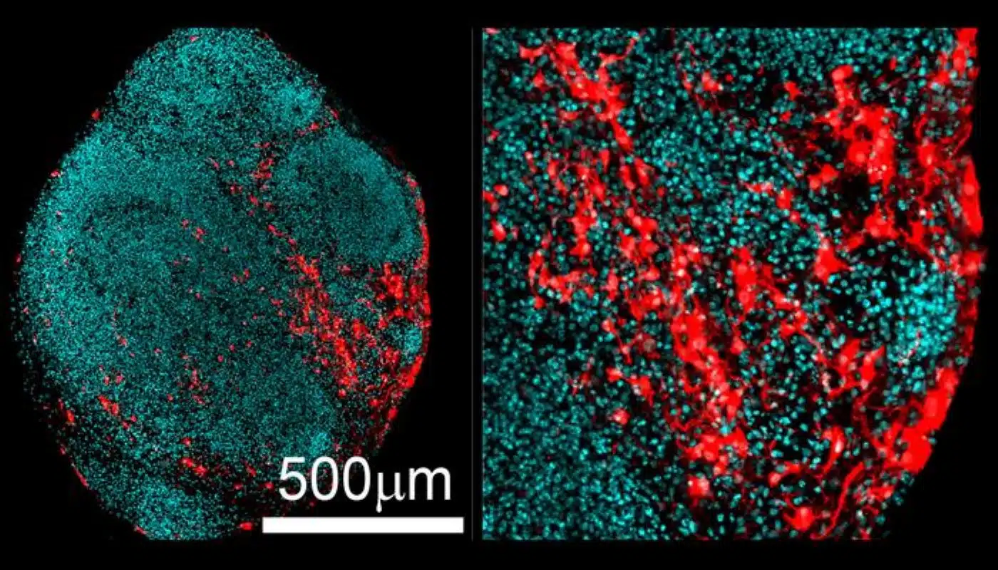
Now, a study published in Nature by researchers at the University of California, Berkeley, overturns this model. By growing stem-cell-derived human brain organoids for up to a year, the team discovered that hyperreactive astrocytes, the star-shaped glial cells that support neuronal networks, are born in a diseased, inflammatory state and directly fuel the development of tuberous lesions.
“As soon as these astrocytes are born, they are reactive and look like they’ve been triggered into a disease state. This is arising as a primary result of the mutation,” said senior author Helen Bateup, professor of neuroscience and of molecular and cell biology at UC Berkeley and a Weill Neurohub Investigator.
“So now we can rethink the disease pathophysiology. It’s not necessarily the case that the neurons are the only cause of seizure activity and the glia become involved later. It could be the other way around, or it could be that both cell types contribute to seizures and epilepsy.”
Recreating the Newborn Brain in a Dish
TSC develops through a two-hit genetic mechanism: a child inherits an altered copy of either TSC1 or TSC2 and subsequently acquires a somatic mutation in the second copy, shutting down critical brakes on the mTOR pathway, a master regulator of cell growth and metabolism.
Investigating these events has historically been hampered by animal models, which fail to mirror the full spectrum of human clinical symptoms. While 3D human brain organoids overcome many of these limitations, human glial development proceeds at a deliberate pace. Radial progenitor cells generate billions of neurons before finally switching to glial production during perinatal and early postnatal life.
To capture this developmental window, the Berkeley team nurtured their organoids in nutrient baths for over nine months to a full year.
“The astrocytes don’t really acquire their mature properties in humans until around perinatal or early postnatal life, so we grow these organoids for a long time,” Bateup explained. “Which people think is crazy. But developing the organoid system was a big advance in our ability to more closely mimic patient brain phenotypes. We now have a much better, or really the only robust model to study tuber cell development.”
In organoids engineered with TSC2 mutations, the radial progenitor cells prematurely produced abnormal, inflammatory astrocytes when they should have been generating healthy neurons.
Shared Signatures with Neurodegeneration
Using single-cell transcriptomics, the investigators mapped the gene expression profiles of individual cells within the organoids. The analysis demonstrated that the hyperreactive astrocytes overexpressed many of the same inflammatory genes seen in neurodegenerative conditions like Alzheimer’s disease.
To confirm these findings outside the laboratory model, the researchers examined surgically resected tuber tissue from 10 pediatric patients with TSC, discovering the identical abnormal protein expression profiles.
Toward Targeted Glial Therapies
Currently, treatment options for mTOR-driven epilepsies, a family of at least 14 genetic conditions known as mTORopathies, rely heavily on systemic mTOR inhibitors such as rapamycin derivatives (rapalogs). While these drugs can decrease seizure frequency, they disrupt vital metabolic, immune, and cellular processes body-wide, causing significant side effects.
Pinpointing astrocytes as the initial culprit opens a more focused therapeutic path: repurposing existing anti-inflammatory and immunosuppressive agents to silence reactive glial signaling without shutting down systemic mTOR pathways.
“If it’s really glia-driven and there’s all these angry cells causing problems, how much can we fix by just suppressing that?” Bateup noted. “Can you calm down the glia and ideally bring them back to a homeostatic state, or if that’s not possible, just shut off their ability to cause damage to the surrounding cells? I think that’s feasible.”
Funding: The work was funded by the National Institute of Neurological Disorders and Stroke (R01NS097823), a Siebel Stem Cell Center Seed grant and a Chan Zuckerberg Biohub investigator award. Thomas Li and John Blair are co-first authors of the paper. Other co-authors are project scientist Taesun Yoo and molecular and cell biology Professor Dirk Hockemeyer of Berkeley and neurosurgeon Gerald Grant and pediatric neurologist Brenda Porter of Stanford University.
Editorial Notes:
- This article was edited by a Neuroscience News editor.
- Journal paper reviewed in full.
- Additional context added by our staff.
About this Epilepsy Research:
- Media Contact: Robert Sanders
- Source: University of California – Berkeley
- Image Credit: Image credited to Helen Bateup/UC Berkeley
- Original Research is Open Access: Nature Communications (September 23, 2026). “mTORC1 drives cell-autonomous astrocyte reactivity in Tuberous Sclerosis.” Authors: Thomas L. Li, John D. Blair, Taesun Yoo, Gerald A. Grant, Dirk Hockemeyer, Brenda E. Porter & Helen S. Bateup.
- DOI: 10.1038/s41586-026-11054-w
Abstract
mTORC1 drives cell-autonomous astrocyte reactivity in Tuberous Sclerosis
Tuberous sclerosis complex (TSC) is a genetic neurodevelopmental disorder characterized by focal brain malformations called cortical tubers, which are associated with severe, intractable epilepsy. Tubers are thought to result from somatic second-hit mutations that disrupt the TSC1 or TSC2 genes in neural progenitor cells, driving hyperactive mTORC1 signalling.
Glial abnormalities are commonly observed in tubers; however, whether glia actively cause disease or merely result from chronic seizure activity has remained unclear. To address this question, we used human brain organoid models to track the developmental fate of mutated progenitor cells in the absence of seizures.
Here we show, using single-cell transcriptomics and cyclic immunostaining across human brain organoids and resected tuber tissue from patients, that loss of TSC2 biases neural progenitors to differentiate into enlarged, pro-inflammatory reactive astrocytes in a cell autonomous manner.
These mutant astrocytes show downregulated glutamate transporter expression, increased inflammatory cytokine secretion and elevated expression of neurodegenerative disease risk genes such as APOE and CLU.
Our findings demonstrate that reactive astrocytes emerge as a primary consequence of TSC2 loss. These results implicate glial dysfunction as a driver of TSC pathogenesis and highlight reactive astrocytes as potential therapeutic targets for TSC-related neuropathology.


