Summary: A new study reveals a mechanism that may explain the link between sound input and cognitive function in the developing brain.
Source: University of Maryland.
Some expectant parents play classical music for their unborn babies, hoping to boost their children’s cognitive capacity later in life. While some research supported a link between prenatal sound exposure and improved brain function, scientists had not identified any structures responsible for this link in the developing brain.
A new study led by University of Maryland neuroscientists is the first to identify a mechanism that could explain such an early link between sound input and cognitive function, often called the “Mozart effect.” Working with an animal model, the researchers found that a type of cell present in the brain’s primary processing area during early development, long thought to form structural scaffolding with no role in transmitting sensory information, may conduct such signals after all.
The results, which could have implications for the early diagnosis of autism and other cognitive deficits, were published in the online early edition of the Proceedings of the National Academy of Sciences on November 6, 2017.
“Previous research documented brain activity in response to sound during early developmental phases, but it was hard to determine where in the brain these signals were coming from,” said Patrick Kanold, a professor of biology at UMD and the senior author of the research paper. “Our study is the first to measure these signals in an important cell type in the brain, providing important new insights into early sensory development in mammals.”
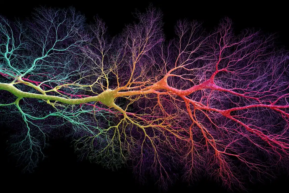
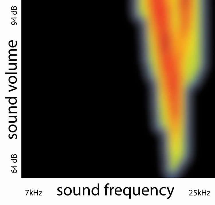
Working with young ferrets, Kanold and his team directly observed sound-induced nerve impulses in subplate neurons for the first time. During development, subplate neurons are among the first neurons to form in the cerebral cortex–the outer part of the mammalian brain that controls perception, memory and, in humans, higher functions such as language and abstract reasoning. Subplate neurons help guide the formation of neural circuits, in the same way that a temporary scaffolding helps a construction crew build walls and install windows on a new building.
Much like construction scaffolding, the role of subplate neurons is thought to be temporary. Once the brain’s permanent neural circuits form, most of the subplate neurons die off and disappear. According to Kanold, researchers assumed that subplate neurons had no role in transmitting sensory information, given their temporary structural role.
Conventional wisdom suggested that mammalian brains transmit their first sensory signals in response to sound after the thalamus fully connects to the cerebral cortex. In many mammals used for research, the connection of the thalamus and the cortex also coincides with the opening of the ear canals, which allows sounds to activate the inner ear. This coincident timing provided further support for the traditional model of when sound processing begins in the brain.
However, researchers had struggled to reconcile this conventional model with observations of sound-induced brain activity much earlier in the developmental process. Until his group directly measured the response of subplate neurons to sound, Kanold said, the phenomenon had largely been overlooked.
“Our work is the first to suggest that subplate neurons do more than bridge the gap between the thalamus and the cortex, forming the structure for future circuits,” Kanold said. “They form a functional scaffolding that actually processes and transmits information before other cortical circuits are activated. It is likely that subplate neurons help determine the early functional organization of the cortex in addition to structural organization.”
By identifying a source of early sensory nerve signals, the current study could lead to new ways to diagnose autism and other cognitive deficits that emerge early in development. Early diagnosis is an important first step toward early intervention and treatment, Kanold noted.
“Now that we know subplate neurons are transmitting sensory input, we can begin to study their functional role in development in more detail,” Kanold said. “What is the role of sensory experience at this early stage? How might defects in subplate neurons correlate with cognitive deficits and conditions like autism? There are so many new possibilities for future research.”
Kanold’s findings are already drawing interest from researchers who study sensory development in humans. Rhodri Cusack, a professor of cognitive neuroscience at Trinity College Dublin, in Ireland, noted that the results could have implications for the care of premature infants.
“This paper shows that our sensory systems are shaped by the environment from a very early age,” Cusack said. “In human infants, this includes the third trimester, when many preterm infants spend time in a neonatal intensive care unit. The findings are a call to action to identify enriching environments that can optimize sensory development in this vulnerable population.”
In addition to Kanold, UMD-affiliated co-authors of the research paper include former graduate student Jessica Wess (Ph.D. ’17, neuroscience and cognitive science) and former postdoctoral researchers Amal Isaiah and Paul Watkins.
The research paper, “Subplate neurons are the first cortical neurons to respond to sensory stimuli,” Jessica Wess, Amal Isaiah, Paul Watkins and Patrick Kanold, was published November 6, 2017 in the online early edition of the Proceedings of the National Academy of Sciences.
Funding: This work was supported by the National Institutes of Health (Award No. R01DC009607) and the Alfred P. Sloan Foundation. The content of this article does not necessarily reflect the views of these organizations.
Source: Matthew Wright – University of Maryland
Publisher: Organized by NeuroscienceNews.com.
Image Source: NeuroscienceNews.com image is credited to Patrick Kanold.
Original Research: Abstract for “Subplate neurons are the first cortical neurons to respond to sensory stimuli” by Jessica M. Wess, Amal Isaiah, Paul V. Watkins, and Patrick O. Kanold in PNAS. Published online November 7 2017 doi:10.1073/pnas.1710793114
[cbtabs][cbtab title=”MLA”]University of Maryland “Source of Early Brain Activity Identified.” NeuroscienceNews. NeuroscienceNews, 7 November 2017.
<https://neurosciencenews.com/early-brain-activity-source-7888/>.[/cbtab][cbtab title=”APA”]University of Maryland (2017, November 7). Source of Early Brain Activity Identified. NeuroscienceNews. Retrieved November 7, 2017 from https://neurosciencenews.com/early-brain-activity-source-7888/[/cbtab][cbtab title=”Chicago”]University of Maryland “Source of Early Brain Activity Identified.” https://neurosciencenews.com/early-brain-activity-source-7888/ (accessed November 7, 2017).[/cbtab][/cbtabs]
Abstract
Subplate neurons are the first cortical neurons to respond to sensory stimuli
In utero experience, such as maternal speech in humans, can shape later perception, although the underlying cortical substrate is unknown. In adult mammals, ascending thalamocortical projections target layer 4, and the onset of sensory responses in the cortex is thought to be dependent on the onset of thalamocortical transmission to layer 4 as well as the ear and eye opening. In developing animals, thalamic fibers do not target layer 4 but instead target subplate neurons deep in the developing white matter. We investigated if subplate neurons respond to sensory stimuli. Using electrophysiological recordings in young ferrets, we show that auditory cortex neurons respond to sound at very young ages, even before the opening of the ears. Single unit recordings showed that auditory responses emerged first in cortical subplate neurons. Subsequently, responses appeared in the future thalamocortical input layer 4, and sound-evoked spike latencies were longer in layer 4 than in subplate, consistent with the known relay of thalamic information to layer 4 by subplate neurons. Electrode array recordings show that early auditory responses demonstrate a nascent topographic organization, suggesting that topographic maps emerge before the onset of spiking responses in layer 4. Together our results show that sound-evoked activity and topographic organization of the cortex emerge earlier and in a different layer than previously thought. Thus, early sound experience can activate and potentially sculpt subplate circuits before permanent thalamocortical circuits to layer 4 are present, and disruption of this early sensory activity could be utilized for early diagnosis of developmental disorders.
“Subplate neurons are the first cortical neurons to respond to sensory stimuli” by Jessica M. Wess, Amal Isaiah, Paul V. Watkins, and Patrick O. Kanold in PNAS. Published online November 7 2017 doi:10.1073/pnas.1710793114