

Summary: A new study questions whether a child’s brain thins as they get older. Researchers say the cortex does not thin during childhood but instead becomes more myelinated.
Source: Max Planck Institute
Using state-of-the-art brain imaging techniques, Vaidehi Natu at Stanford University in California, along with her colleagues from the Max Planck Institute for Human Cognitive and Brain Sciences in Leipzig, Germany, provide striking results that suggest the brains of children likely do not thin as much as expected. Rather, it seems there is an increase of myelin, which is the fatty sheath insulating nerve fibers.
Stepping back a bit, studies have shown repeatedly that certain regions of the cerebral cortex (the outer-most layer of the brain) get thinner as children develop. And at a mere 3mm thick, on average, studies have reported that kids can apparently lose close to 1mm of gray matter by adulthood. Various hypotheses have been put forth to explain these huge losses. For example, it is established that gray matter cells and their connections can be naturally ‘pruned’, presumably to promote a more efficient brain. So perhaps extensive pruning in young brains could explain the thinning. Alternatively, we know that our brains expand during development. Perhaps the cortex gets stretched in the process? The new research certainly does not rule out these processes, and in fact finds evidence for the latter. However, the new work does suggest that a prominent change has gone undetected, due to limitations of prior measurements.
More specifically, the present study shows that when measured with quantitative MRI (or qMRI), it appears that young brains are actually becoming more myelinated. That’s a good thing, but it may be messing up estimates of cortical thickness (the gray matter). Myelin is the ‘white’ in white matter. It’s a fatty sheath that insulates many nerve fibers and allows faster neurotransmission. The problem is, measurements of gray matter thickness critically depend on detecting the border between white and gray matter. As Natu and Kirilina have found out, this limit can be obscured and cortical thickness underestimated if myelination increases during development. The researchers obtained these results by examining adults and children using state-of-the-art quantitative MRI techniques.
Evgeniya Kirilina, who is working in the department of Neurophysics at MPI CBS, offers a word of caution. ‘The fact that the cortex thins during development is well established, even with histological methods. We are not claiming that thinning does not occur. But estimates may be off in some cases due to concurrent myelination.’
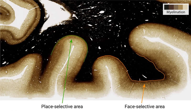
The team was actually looking at three specialized patches of brain in higher visual cortex. Despite their close proximity, each showed a unique developmental pattern, underlining the need for cautious interpretation. The face and word recognition areas showed the myelination effect described above, whereas the place recognition area showed apparent thinning but no indication of myelination. Instead, it seemed to structurally change, stretching over time. Highlighting the important link between structure and function, the differences in myelination of these functionally specialized regions were confirmed in post mortem brains of adults, using both ultra-high field MRI and histology.
The implications of these new findings are quite broad. Decades of work will need to be revisited and assessed for accuracy. For example, there is a rich literature suggesting that the thickness of the cortex changes when learning new skills. It will now need to be determined whether myelination also plays a role. Further, degradation of myelin can lead to debilitating diseases. This is exactly what occurs in Multiple Sclerosis. More accurate measurement techniques like qMRI promise to improve our detection, monitoring, and treatment of such conditions.
Source:
Max Planck Institute
Media Contacts:
Evgeniya Kirilina – Max Planck Institute
Image Source:
The image is credited to MPI CBS.
Original Research: Open access
“Apparent thinning of human visual cortex during childhood is associated with myelination”. Vaidehi S. Natu, Jesse Gomez, Michael Barnett, Brianna Jeska, Evgeniya Kirilina, Carsten Jaeger, Zonglei Zhen, Siobhan Cox, Kevin S. Weiner, Nikolaus Weiskopf, and Kalanit Grill-Spector.
PNAS doi:10.1073/pnas.1904931116.
Abstract
Apparent thinning of human visual cortex during childhood is associated with myelination
Human cortex appears to thin during childhood development. However, the underlying microstructural mechanisms are unknown. Using functional magnetic resonance imaging (fMRI), quantitative MRI (qMRI), and diffusion MRI (dMRI) in children and adults, we tested what quantitative changes occur to gray and white matter in ventral temporal cortex (VTC) from childhood to adulthood, and how these changes relate to cortical thinning. T1 relaxation time from qMRI and mean diffusivity (MD) from dMRI provide independent and complementary measurements of microstructural properties of gray and white matter tissue. In face- and character-selective regions in lateral VTC, T1 and MD decreased from age 5 to adulthood in mid and deep cortex, as well as in their adjacent white matter. T1 reduction also occurred longitudinally in children’s brain regions. T1 and MD decreases 1) were consistent with tissue growth related to myelination, which we verified with adult histological myelin stains, and 2) were correlated with apparent cortical thinning. In contrast, in place-selective cortex in medial VTC, we found no development of T1 or MD after age 5, and thickness was related to cortical morphology. These findings suggest that lateral VTC likely becomes more myelinated from childhood to adulthood, affecting the contrast of MR images and, in turn, the apparent gray–white boundary. These findings are important because they suggest that VTC does not thin during childhood but instead gets more myelinated. Our data have broad ramifications for understanding both typical and atypical brain development using advanced in vivo quantitative measurements and clinical conditions implicating myelin.


