Summary: Researchers have identified a mechanism that occurs within the CA3 region of the hippocampus that appears to be responsible for a common type of age-related memory loss.
Source: Johns Hopkins University
Working with rats, neuroscientists at Johns Hopkins University have pinpointed a mechanism in the brain responsible for a common type of age-related memory loss.
The work, published today in Current Biology, sheds light on the workings of aging brains and may deepen our understanding of Alzheimer’s disease and similar disorders in humans.
“We’re trying to understand normal memory and why a part of the brain called the hippocampus is so critical for normal memory,” said senior author James Knierim, a professor at the university’s Zanvyl Krieger Mind/Brain Institute. “But also with many memory disorders, something is going wrong with this area.”
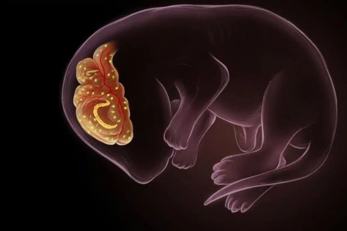
Neuroscientists know that neurons in the hippocampus, located deep in the brain’s temporal lobe, are responsible for a complementary pair of memory functions called pattern separation and pattern completion. These functions occur in a gradient across a tiny region of the hippocampus called CA3.
When those functions swing out of balance, memory becomes impaired, causing symptoms like forgetfulness or repeating oneself. The Johns Hopkins team discovered that as the brain ages, this imbalance may be caused by the CA3 gradient disappearing; the pattern separation function fades away, and the pattern completion function takes over.
Neurons responsible for pattern separation are typically more prevalent in the proximal region of the CA3 area, while those responsible for pattern completion are prevalent in the distal region, said lead author Heekyung Lee, an assistant research scientist at the Mind/Brain Institute, With aging, neural activity in the proximal region becomes overactive, and the interplay between the two regions becomes abnormal, creating a dominance in pattern completion.
In normal brains, pattern separation and pattern completion work hand-in-hand to sort and make sense of perceptions and experiences, from the most basic to the highly complex.
If you visit a restaurant with your family and a month later you visit the same restaurant with friends, you should be able to recognize that it was the same restaurant, even though some details have changed—this is pattern completion.
But you also need to remember which conversation happened when, so you do not confuse the two experiences—this is pattern separation.
When pattern separation disappears, pattern completion overpowers the process. With your brain focusing on the common experience of the restaurant to the exclusion of the details of the separate visits, you might remember a conversation about a trip to Italy during one visit, but mistake who was talking.
“We all make these mistakes, but they just tend to get worse with aging,” Knierim said.
In experiments the researchers compared young rats with unimpaired memories to older rats with unimpaired memories and older rats with impaired memories.
While the older rats with unimpaired memories performed water maze tasks as well as young rats, the neurons in the CA3 regions of their hippocampi were already beginning to favor pattern completion at the expense of pattern separation.

Since that physiological finding had not shown up in their behavior, the researchers concluded that something was allowing the rats to compensate for the deficit.
That finding is echoed in humans who remain surprisingly sharp into their older years, the researchers say. So pinpointing the memory loss mechanism could lay the groundwork for learning what prevents memory impairment in some humans, and therefore how to prevent or delay cognitive decline in the elderly.
“If we can understand better what these compensatory mechanisms are, then maybe we can help prevent cognitive decline with aging,” Knierim said. “Or, if we can’t stop it, maybe we can enhance other parts of the brain to compensate for the losses that are occurring.”
Other senior authors of the paper were Michela Gallagher, Krieger-Eisenhower Professor of Psychology and Neuroscience at Johns Hopkins, and Scott Zeger, professor of biostatistics at Johns Hopkins’ Bloomberg School of Public Health. Gallagher’s lab previously demonstrated that the anti-epilepsy drug Levetiracetam improves memory performance by reducing hyperactivity in the hippocampus. So Lee also speculates that this new, more specific information about how memory impairment occurs might allow scientists to better aim such drugs toward the deficits in the future.
“It would give us better control of where we could possibly target the deficits that we see,” she said.
About this aging and memory research news
Author: Press Office
Source: Johns Hopkins University
Contact: Press Office – Johns Hopkins University
Image: The image is in the public domain
Original Research: Closed access.
“Loss of functional heterogeneity along the CA3 transverse axis in aging” by Heekyung Lee et al. Current Biology
Abstract
Loss of functional heterogeneity along the CA3 transverse axis in aging
Highlights
- Young (Y) rats show transition from pattern separation to pattern completion in CA3
- Aged memory-impaired (AI) rats show pattern completion in proximal and distal CA3
- AI rats can orthogonalize representations in two spatially, distinct environments
- Aged memory-unimpaired rats show trends that are intermediate between Y and AI rats
Summary
Age-related deficits in pattern separation have been postulated to bias the output of hippocampal memory processing toward pattern completion, which can cause deficits in accurate memory retrieval.
Although the CA3 region of the hippocampus is often conceptualized as a homogeneous network involved in pattern completion, growing evidence demonstrates a functional gradient in CA3 along the transverse axis, as pattern-separated outputs (dominant in the more proximal CA3) transition to pattern-completed outputs (dominant in the more distal CA3).
We examined the neural representations along the CA3 transverse axis in young (Y), aged memory-unimpaired (AU), and aged memory-impaired (AI) rats when different changes were made to the environment.
Functional heterogeneity in CA3 was observed in Y and AU rats when the environmental similarity was high (altered cues or altered environment shapes in the same room), with more orthogonalized representations in proximal CA3 than in distal CA3.
In contrast, AI rats showed reduced orthogonalization in proximal CA3 but showed normal (i.e., generalized) representations in distal CA3, with little evidence of a functional gradient.
Under experimental conditions when the environmental similarity was low (different rooms), representations in proximal and distal CA3 remapped in all rats, showing that CA3 of AI rats is able to encode distinctive representations for inputs with greater dissimilarity.
These experiments support the hypotheses that the age-related bias toward hippocampal pattern completion is due to the loss in AI rats of the normal transition from pattern separation to pattern completion along the CA3 transverse axis.